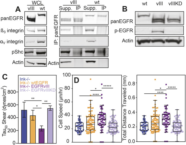
Fig. 4.
Adhesion strength is modulated by kinase-dependent mechanism(s). (A) Western blots of whole-cell lysate (WCL; left) confirm genotypes and expression patterns of integrins αV and β3. Additional blots (right) for EGFRvIII and wtEGFR show the results of EGFR immunoprecipitation with supernatant (Supp.) and pulldown (IP) lanes for the integrins αV and β3, phosphorylated SHC (pShc) and actin. (B) Western blot for total EGFR (pan-EGFR), phosphorylated EGFR (pEGFR) and actin for wtEGFR (wt), EGFRvIII (vIII) and EGFRvIIIKD (vIIIKD). (C) Adhesion strength, i.e. τ50, plotted for the different genotypes as indicated. *P<0.05 and **P<0.01 assessed by paired Student’s t-test (n=4 for each genotype). (D) Cell speed and total migration distance plotted for the same genotypes as listed in panel C. *P<0.05; ****P<0.0001; *****p<0.00001; assessed by paired Student’s t-test (n=4 biological replicates, analyzing 39, 88, 65 and 93 cells for Ink−/−, wtEGFR, EGFRvIII and EGFRvIIIKD cells, respectively).

