Fig. 5.
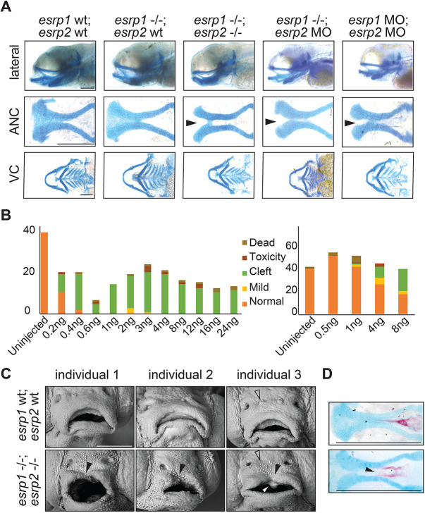
esrp1/2 double mutants display a cleft lip and palate. (A) Alcian Blue staining of 4 dpf zebrafish. Representative images of WT, esrp1 CRISPR mutant (esrp1−/−) and esrp1/2 double CRISPR mutant (esrp1−/−; esrp2−/−), as well as esrp1 CRISPR mutant treated with esrp2 morpholino and WT treated with esrp1 and esrp2 morpholino (esrp1 MO, esrp2 MO). Flat-mount images of the anterior neurocranium (ANC) show a cleft (arrowheads) between the median element and lateral element of the ANC when both esrp1 and esrp2 function were disrupted. Lateral images and flat-mount images of the ventral cartilage (VC) show only subtle changes in morphology between WT and esrp1/2−/− zebrafish. (B) Morphant phenotypes observed over a range of esrp1 and esrp2 MO doses. Single esrp2 MO injections in the esrp1−/− background achieves nearly 100% phenotype penetrance, even at very low MO doses. (C) SEM of 5 dpf zebrafish showing discontinuous upper lip (filled arrowheads) in the esrp1/2 double CRISPR mutant as well as absent preoptic cranial neuromasts (open arrowheads) and abnormal keratinocyte morphology. The white arrowhead indicates an aberrant cell mass. (D) Representative images of Alizarin Red/Alcian Blue staining of 9 dpf esrp1/2 double CRISPR mutant zebrafish and WT clutch-mate controls. Esrp1/2 ablation causes abnormal morphology of the mineralizing parasphenoid bone; the bone appears wider and with a cleft (arrowhead). Scale bars: 150 µm (A,D); 100 µm (C).

