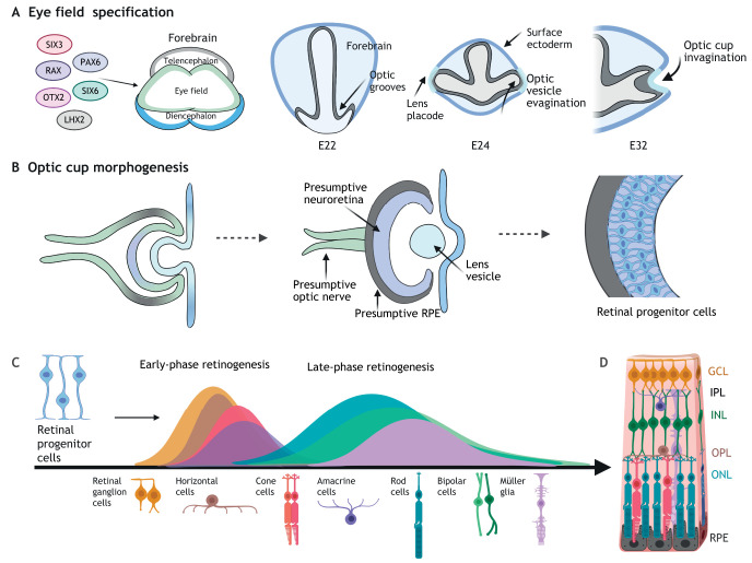
Fig. 1.
Building the retina: eye field specification, optic cup morphogenesis and retinal cell differentiation. (A) In vivo, the eye field transcription factors SIX3, RAX, PAX6, OTX2, SIX6 and LHX2 specify the presumptive eye field in the diencephalon of the developing forebrain. By human embryonic day 22 (E22), indents (optic grooves) form in the neural fold, bilaterally to the diencephalon. The optic grooves evaginate by E24 to form the optic vesicles, which protrude to contact the overlying surface ectoderm at the site of the presumptive lens (lens placode). Invagination of the optic vesicle forms the bilayered optic cup by E32. (B) The inner layer of the optic cup specifies the presumptive neuroretina, whereas the outer layer specifies the presumptive retinal pigment epithelium (RPE). The optic vesicles remain connected to the forebrain via the optic stalk, a hollow connection that closes to form the presumptive optic nerve. The lens vesicle pinches off the surface ectoderm. In the presumptive neuroretina, multipotent retinal progenitor cells (RPCs) begin to differentiate into retinal cell types. (C) Differentiation of the seven main retinal cell types from RPCs proceeds sequentially in waves, with retinal ganglion cells, horizontal cells, cone photoreceptor cells and amacrine cells formed in an early retinogenesis wave, followed by the overlapping late-phase generation of rod photoreceptor, bipolar and Müller glia cells. (D) These cell types populate the multi-layered retina from the basal-most ganglion cell layer (GCL), inner plexiform layer (IPL), inner nuclear layer (INL), outer plexiform layer (OPL) and apical-most outer nuclear layer (ONL), where photoreceptors lie adjacent to the RPE.

