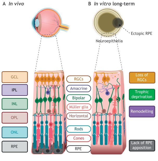
Fig. 4.
Modelling retinal layers in vitro. (A) In vivo, the seven main neuroretinal cell types populate the layers of the retina with retinal pigmented epithelium (RPE) next to the outer nuclear layer (ONL). Interneurons synapse with photoreceptors in the outer plexiform layer (OPL) and retinal ganglion cells (RGCs) in the inner plexiform layer (IPL) to relay signals to the brain. (B) In vitro, retinal organoids develop multiple layers and cell types, but RGCs are progressively lost in long-term culture, possibly owing to lack of neurotrophic factors or other ocular structures. Subsequently, interneuron cells are lost and remodelling occurs, possibly owing to trophic deprivation caused by loss of synaptic partner RGCs. In retinal organoids, RPE, a major source of diffusible factors, forms in adjacent clumps rather than juxtaposed to the ONL.

