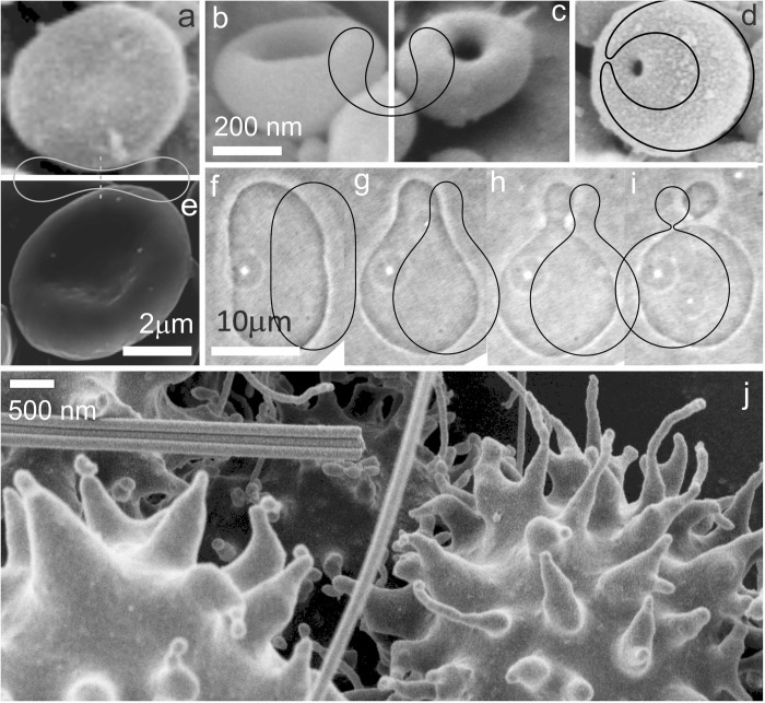
Fig 3. Comparison between experimental and theoretical shapes.
a-d: The transformation of a discocyte into a stomatocyte as represented by the shapes observed in cellular nanovesicles, and the corresponding contours of the calculated shapes obtained by minimization of the membrane free energy. e: Discocyte shape of an erythrocyte complying with discocyte shape of cellular nanovesicle and with the calculated shape. f-i: The transformation of the outward bud in giant phospholipid vesicles and the corresponding result of the theoretical description obtained by minimization of the membrane free energy. The parameters of the calculated shapes are hm = dm = 0, (a,e): v = 0.6, <h> = 1.040, <d> = 1.812, (b,c): v = 0.6, <h> = 0.650, <d> = 1.167, (d): v = 0.6, <h> = 0.435, <d> = 0.235, (f): v = 0.9, <h> = 1.050, <d> = 0.729, (g): v = 0.9, <h> = 1.105, <d> = 0.697, (h): v = 0.9, <h> = 1.155, <d> = 0.577, (i): v = 0.9, <h> = 1.240, <d> = 0.163.

