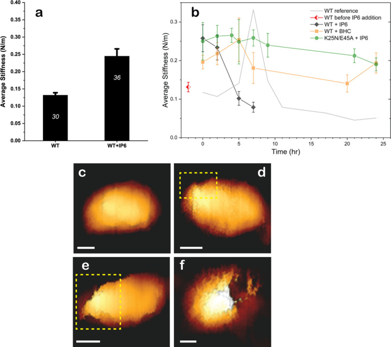
Fig 4. Morphologies and mechanical properties of HIV-1 capsids from AFM.

(a) The average stiffness of isolated WT HIV-1 capsids in the absence (n = 30) and presence (n = 36) of IP6. Error bars indicate SEM. Statistical significance was confirmed using the Mann–Whitney U test for p < 0.05. (b) Average stiffness of HIV-1 capsids treated with 100 μM IP6 (black) or BHC (orange) as a function of time after the addition of dNTPs and MgCl2. The red diamond represents the average stiffness of WT capsids before addition of IP6 or BHC. The initial average stiffness, at time 0, was measured before addition of dNTPs and MgCl2. At each time point, the stiffness of 2 to 4 capsids was measured without stopping the reaction. WT data (taken from [17]) is presented as a gray line for comparison. The error bars represent SEM. (c) A representative cone-shaped capsid (of 20 imaged) prior to addition of dNTP and MgCl2. (d–f) Deformed and damaged capsids visualized after 5 h. For clarity, openings in the capsids are shown within a dashed yellow rectangle. Scale bars, 50 nm. A total of 52 capsids were visualized. (Approximately 400 force-distance curves were obtained from individual capsids). Numerical data for panel A and B can be found in S4 Data. AFM, atomic force microscopy; BHC, benzenehexacarboxylic acid; dNTP, deoxynucleotide triphosphate; IP6, inositol hexakisphosphate; WT, wild-type.
