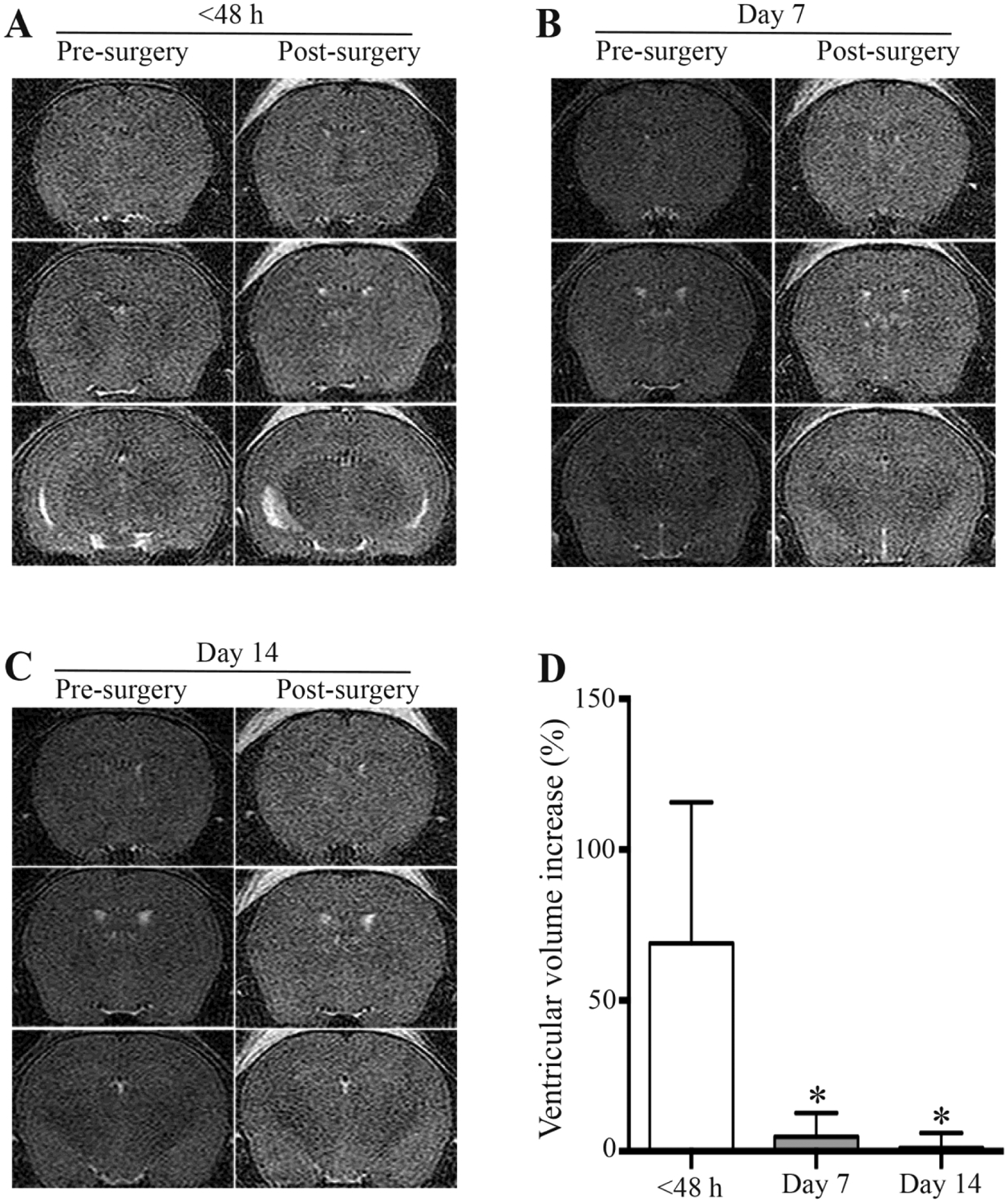
Fig. 3.

Ventricular enlargement after intracerebroventricular (ICV) injection of acellular CSF from SAH patient 1, where the CSF was sampled at different time points after ictus. Examples of MRIs of nude mice prior to and 24 hours after ICV injection of acellular CSF, where the CSF was sampled at (A) <48 hours, (B) day 7 and (C) day 14. (D) Quantification of the degree of ventricular enlargement. Values are means ± SD, n = 7, *p < 0.05 vs. <48 hours.
