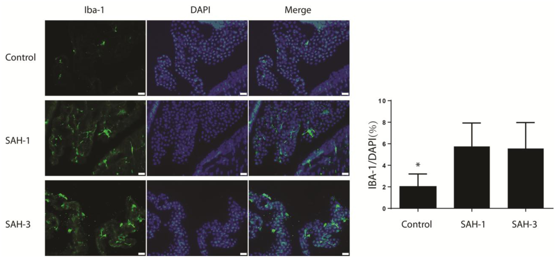
Fig. 6.

Epiplexus cell activation at 24 hours following ICV injection of acellular CSF from a control patient and from SAH patients 1 and 3. Immunohistochemistry for Iba-1 in the choroid plexus. Iba-1 is a macrophage marker that detects epiplexus cells. The same sections were counterstained with DAPI, a nuclear marker, and the merged images are shown. The number of Iba-1 cells was quantified as a percent of all cells as determined by DAPI. Values are means ± SD, n = 7, *p < 0.05 vs. the other groups. Scale bar = 20 μm.
