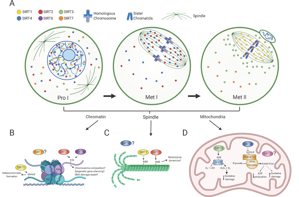
Figure 1:
Sirtuin distribution in oocytes during meiotic divisions and its associated molecular functions. A) Scheme denotes Sirtuin localization in oocytes at prophase I (Pro I) and at Metaphase I and II (Met I, Met II). SIRT1 is mostly nuclear in prophase I-arrested oocytes and on the spindle in Met II-arrested eggs. SIRT2 is found throughout the ooplasm and nucleus in prophase I and relocalizes to the spindle in Met I. SIRT6 and SIRT7 are chromatin-bound at all developmental stages, but SIRT7 is also present to a lesser extent in the ooplasm. SIRT3 and SIRT4 are predominantly nuclear in prophase I. SIRT3 redistributes around the spindle in Met II eggs and SIRT4 reallocates to the spindle in Met I. The cellular distribution of SIRT5 in oocytes is currently unknown. B-D) Molecular functions of Sirtuins during meiotic maturation in chromatin (B), the spindle (C) and mitochondria (D). B) SIRT1 promotes H3K9me3 deposition and heterochromatin formation and SIRT2 and SIRT6 regulates H4K16ac levels with potential implications in chromosome compaction, gene silencing and DNA damage repair signaling. C) SIRT1 and SIRT2 both regulate acetylation of α-tubulin suggesting a potential role in spindle dynamics. D) SIRT3 stimulates SOD2 activity and ROS balance and SIRT4 limits Pyruvate Dehydrogenase (PDH) complex activity leading to reduced ATP production. Solid and dashed arrows denote potential direct and indirect roles, respectively. Created with BioRender.com.

