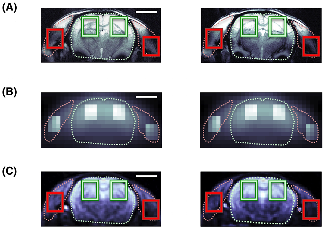
FIGURE 1.

Co-registered rat head images with 5mm scale bar: (A), 1H FLASH structural image of the coronal rat head anatomy. Regions of interest (ROI) for brain tissue (green) are highlighted for each hemisphere and laterally left and right for muscle tissue (i.e. predominantly temporalis lateralis; red); (B), in vivo H217O image acquired within 30 s shortly before the onset of a 17O2 inhalation, with intensity highlighting (2× brighter) of the ROIs for better visualization; and (C), very high resolution H217O enriched post-mortem image acquired after repeated inhalations with ROIs marked as in (A). All slices cover the same FOV at the Z-position of the Bregma. Visualization of brain and muscle (dotted line) based on proton images (A). 17O images in (B,C) are zero-filled (×2) in the spatial dimensions.
