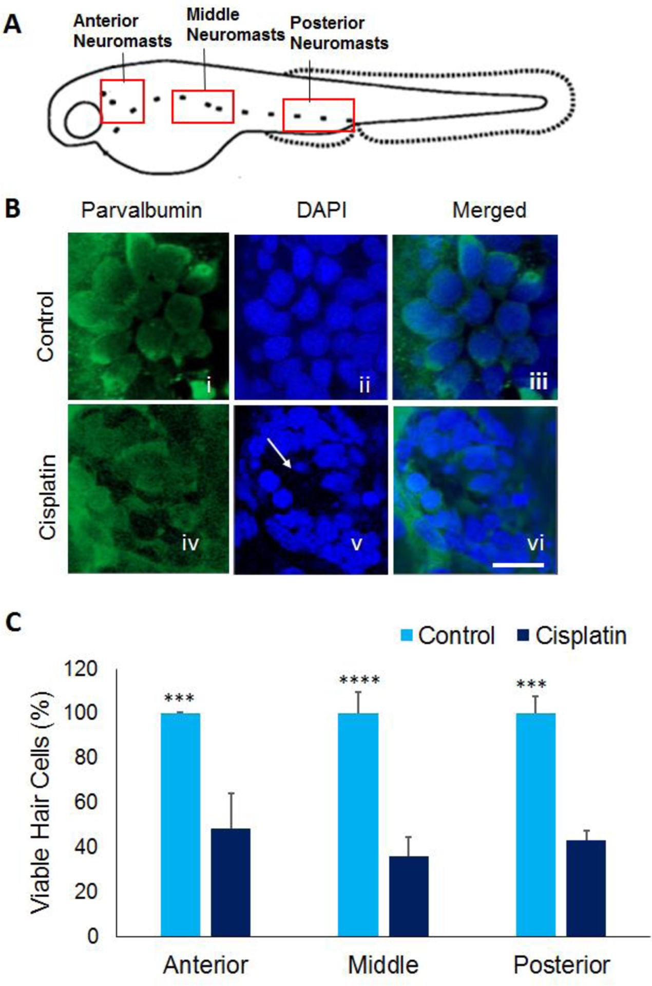
Figure 1: Cisplatin-induced hair cell loss.

(A) Schematic representation of neuromasts in the anterior, middle, and posterior regions of 5dpf zebrafish larvae. (B) Treatment of 5dpf larvae with 1000 μM cisplatin induced hair cell loss in neuromasts located in the anterior, middle, and posterior regions along the lateral line. The hair cells were labelled with parvalbumin (green), a hair cell marker (i and iv), images were captured at 63X magnification, and the hair cells were counted manually. Cisplatin-induced loss of hair cells is evident from lack of intact hair cells and nuclei (white arrow) in panel iv and v, respectively. Scale bar = 5 μm. (C) Quantification of the hair cells indicated that cisplatin treatment induced a significant decrease in hair cell viability in all three regions along the lateral line suggesting the toxic effect of cisplatin in zebrafish hair cells. The results are expressed as mean ± standard deviation, n = 12, (***p<0.001, ****p<0.0001).
