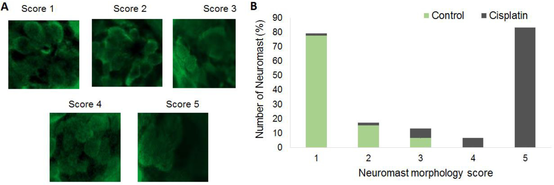
Figure 2: Morphometric analysis of neuromasts.

(A) Representative images indicating the degree of distortion in the morphology of hair cells and neuromasts and their corresponding scores are illustrated. The hair cells were labelled with parvalbumin (green) and the images were captured at 63X magnification. The morphometric analysis indicated that the morphology of hair cells and the neuromasts were distorted after cisplatin treatment. (B) Morphometric scores indicated that more than 77% of the neuromasts in the controls had intact and well-defined morphology. However, 83% of the neuromasts in cisplatin treated larvae received a score of 5 suggesting that the hair cells were dispersed and the neuromasts were distorted, n = 6. Evaluation of the difference in the morphology by Mann-Whitney’s U test indicated that the differences were significant between the control and cisplatin-treated groups (mean scores of control and cisplatin-treated groups were 1 and 5, respectively; U=155; p<0.001).
