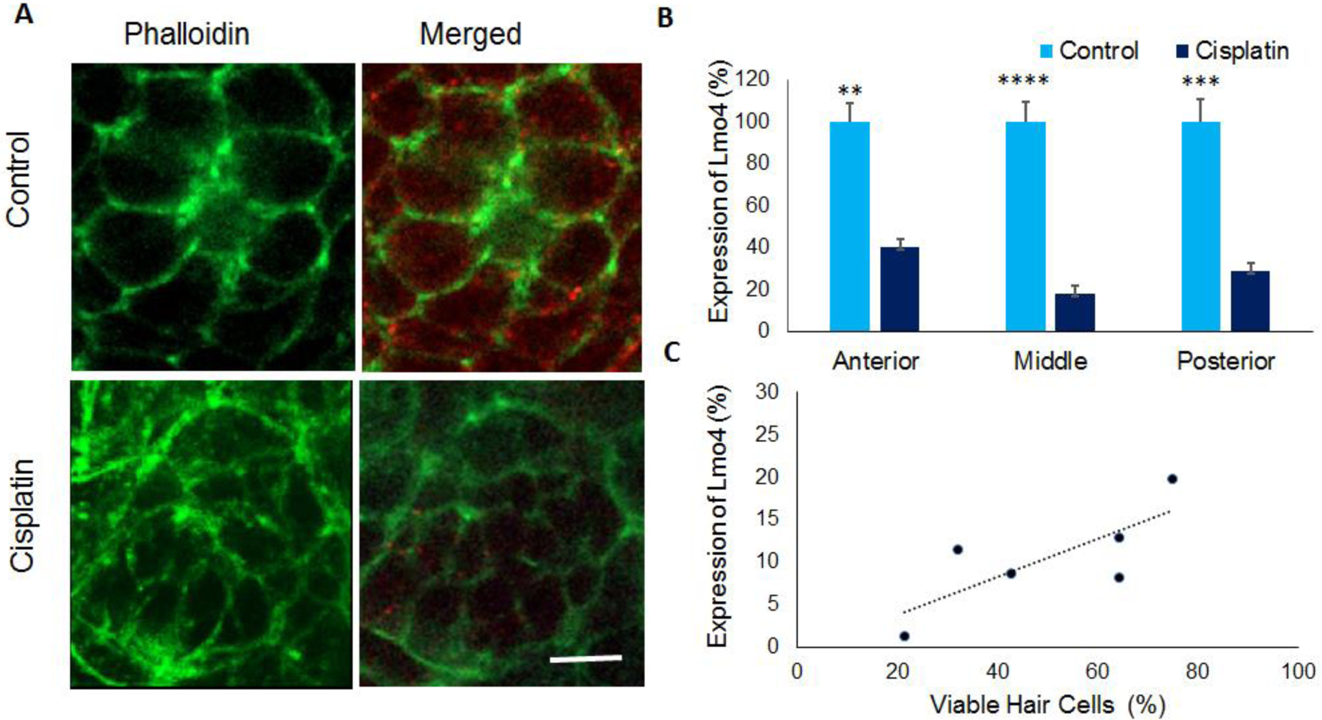
Figure 3: Lmo4 protein expression in zebrafish hair cells.

(A) Immunohistochemistry analysis indicated that cisplatin treatment decreased the expression of Lmo4 protein (red) in the hair cells of 5dpf stage larvae. Phalloidin (green) was used to stain actin in the hair cells. Images are representative of six replicates. Scale bar= 5 μm. (B) Quantification of the staining intensity in neuromasts located in the anterior, middle, and posterior regions indicated a significant decrease in the Lmo4 protein levels in cisplatin treated zebrafish. The results are expressed as mean ± standard error, n = 6, (**p<0.01, ***p<0.001, ****p<0.0001). (C) Analysis of the correlation between cisplatin-induced changes in hair cell viability and Lmo4 protein expression indicated a positive correlation (R2=0.76), n=6.
