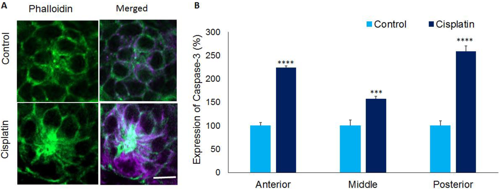
Figure 5: Activated Caspase-3 expression in zebrafish hair cells.

(A) Immunolocalization with anti- Caspase-3 indicated that cisplatin treatment increased the expression of activated Caspase-3 (purple) in the hair cells of 5dpf stage larvae. This suggested cisplatin-induced apoptosis in zebrafish hair cells. Phalloidin (green) was used to stain actin in the hair cells. Images are representative of six replicates. Scale bar= 5 μm. (B) Quantification of the staining intensity in neuromasts located in the anterior, middle, and posterior regions indicated a significant increase in activated Caspase-3 levels in cisplatin treated zebrafish. The results are expressed as mean ± standard error, n = 6, (***p<0.001, ****p<0.0001).
