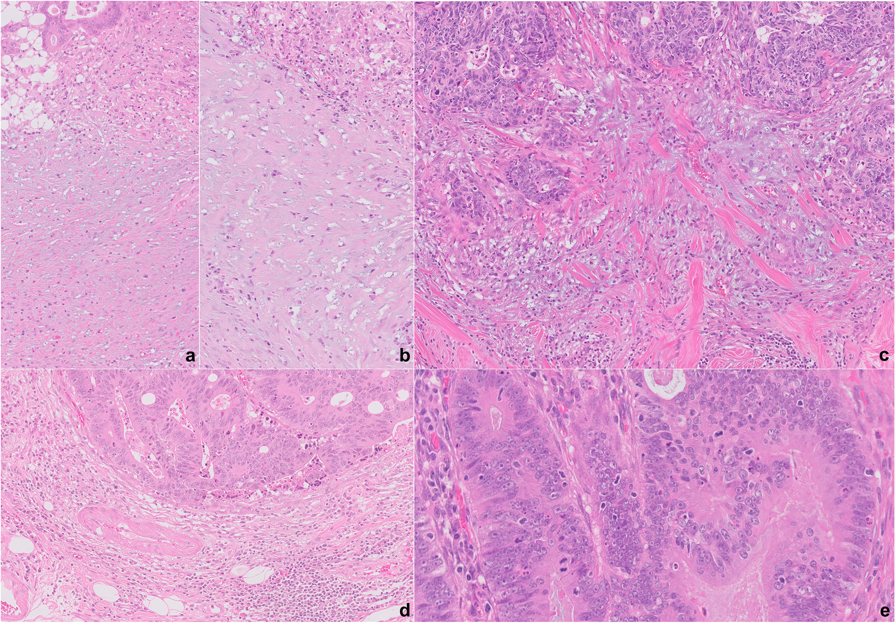
Figure 1.

A–D, Examples of desmoplastic reaction (DR) at the tumour edge. An immature/myxoid DR, extending at least a high-power field (×40) (A,B); intermediate DR, with keloid-like collagen (C); and mature DR, without immature/myxoid stroma or keloid-like collagen (D). Intraepithelial tumour infiltrating lymphocytes, defined as ≥ 5 lymphocytes per high-power field (×40) (E).
