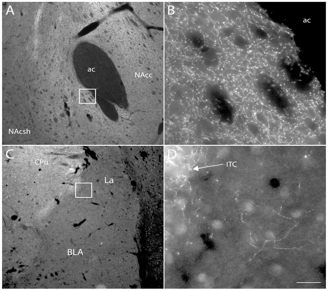
Figure 4. Localization of DAT expression in NAcc and BLA.
Fluorescence photomicrographs depicting immunofluorescence localization of dopamine transporter (DAT) immunoreactivity in the nucleus accumbens (A, B) and basolateral amygdala (C, D) of the rat. Boxes in (A) and (C) are shown at higher magnification in (B) and (D), respectively. DAT-immunoreactive fibers were observed at high density in the nucleus accumbens (B) and dorsal striatum (C), and at much lower density in the BLA (D). DAT-immunoreactive fibers were observed at higher density in the intercalated cell groups (ITC) of the amygdala (C, D) than in the rest of the amygdala. Scale bar = 200 μm (A, C); 20 μm (B, D). ac – anterior commissure; BLA – basolateral amygdala; ITC –intercalated cell group; NAcc – nucleus accumbens core; NAcsh – nucleus accumbens shell.

