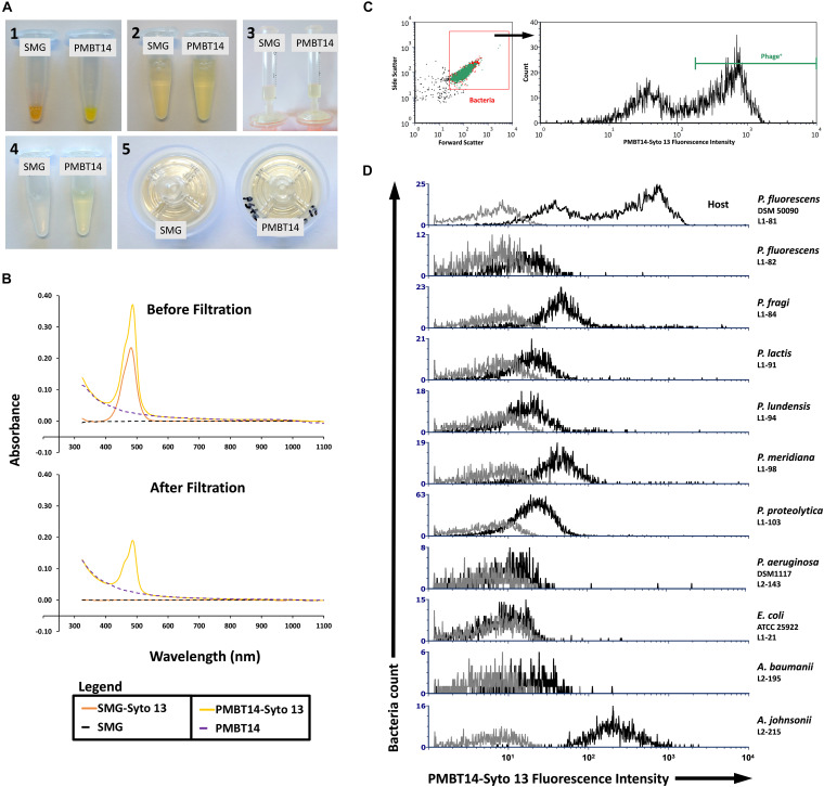
FIGURE 1.
Syto 13 labeling of PMBT14 and its use in staining host bacteria. (A) Process of labeling bacteriophage using Syto 13 by (1) adding 1 μL Syto 13 to 20 μL 1013 PFU/mL bacteriophage (negative control SMG buffer only); (2) equilibrating the staining solution with 980 μL SMG buffer; (3) slowly passing the bacteriophage-Syto 13 mix through a 0.45 μm PES syringe filter; (4) filtrate; (5) retentate. (B) UV-Vis spectrophotometry of SMG buffer or PMBT14 treated with and without Syto 13, before and after filtration through the 0.45 μm filter. (C) Gating strategy for the flow cytometry analysis of bacteria staining with PMBT14-Syto13. Bacteria cells were first crudely gated based on their forward and sideward light scatter (left) and the infected “Phage+” cells were defined as cells that stained very positively with Syto 13 (right). (D) Histograms of PMBT14-Syto13 staining on host L1-81 P. fluorescens as well as a selection of other related and unrelated bacteria (black line). Unstained controls are in gray.

