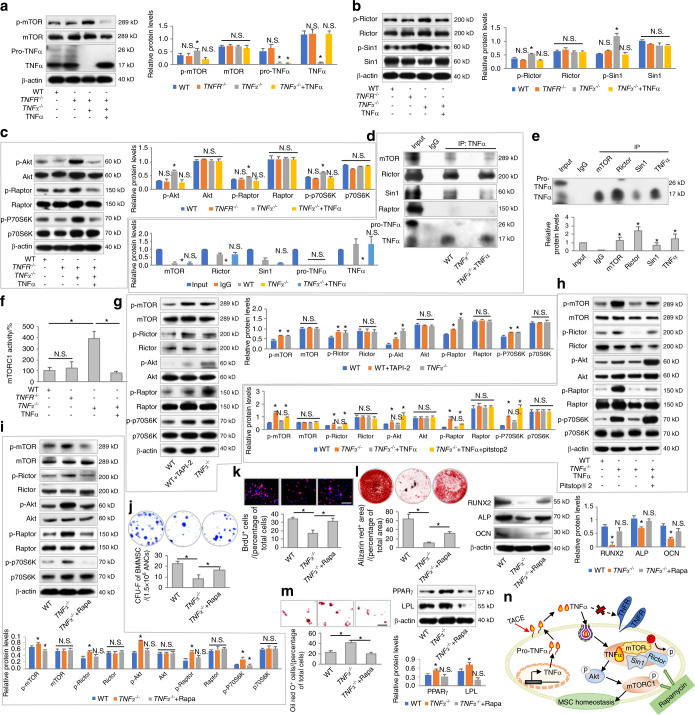
Fig. 3.
Endocytosed TNFα binds to mTORC2 and restricts activation of mTOR signaling for functional regulation of MSCs. a–c Western blot analyses of mTOR signaling in response to TNFR or TNFα deficiency. N = 3. TNFα was added at 1 ng·mL−1. d, e Co-IP analyses of the binding of TNFα to mTOR complex components (N = 3). f Analysis of mTORC1 activity using an ELISA-based assay (N = 3). g–i Western blot analyses of mTOR signaling. N = 3. TAPI-2, TNFα, Pitstop® 2, and rapamycin (Rapa) were added at 120 nmol·L−1, 1 ng·mL−1, 12 μmol·L−1, and 50 nmol·L−1, respectively, in vitro. j–m Functional analyses of MSCs according to CFU, BrdU labeling, and osteogenic and adipogenic differentiation (N = 3). Scale bars = 100 μm. n Diagram showing TNFα binding to mTORC2 and restricting activation of mTOR signaling for functional regulation of MSCs. For quantification of Western blotting, a two-tailed Student’s t test was used for the comparison between the treatment and WT groups. *P < 0.05. N.S. not significant. Data represent the mean ± SD

