
Figure 5 |. MRI findings in haemochromatosis owing to hepcidin deficiency and ferroportin disease.
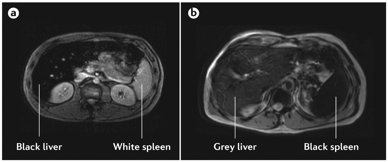
The signal intensity ratio technique with T2-weighted MRI can be used to differentiate patients with haemochromatosis and those with ferroportin disease. a | In patients with hepcidin-deficient haemochromatosis, ‘black’ liver corresponds to a highly iron-overloaded liver, and ‘white’ spleen corresponds to the absence of iron overload. The appearance of the liver and spleen is similar in patients with types 1, 2A, 2B, 3 and 4 haemochromatosis. b | In ferroportin disease, the spleen appears black (highly iron-overloaded) and the liver appears grey (moderately iron-overloaded) or black (highly iron-overloaded) on T2-weighted MRI47.