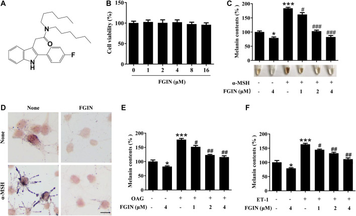
FIGURE 1.
Effect of FGIN-1-27 (FGIN) on melanogenesis in SK-MEL-2 cells. (A) The chemical structure of FGIN-1-27, (B) after incubation of with various concentrations (1–16 μM) of FGIN-1-27 for 48 h, cell viability was determined using MTT assay, (C) SK-MEL-2 cells were treated with FGIN-1-27 in the presence or absence of α-MSH (50 nM) for 48 h. Melanin contents were measured as described in methods. (D) SK-MEL-2 cells were treated with FGIN-1-27 (4 μM) for 48 h and were stained with Masson–Fontana ammoniacal silver stain. Bar = 20 μm (E,F) SK-MEL-2 cells were treated with 200 μM OAG or 10 nM ET-1 in the presence or absence of FGIN-1-27. Melanin contents were measured as described in methods. Data are expressed as the mean ± SD (n = 3). *p < 0.05, ***p < 0.001 vs. non-treated cells. # p < 0.05, ## p < 0.01, ### p < 0.001 vs. α-MSH-, OAG-, or ET-1-treated cells.

