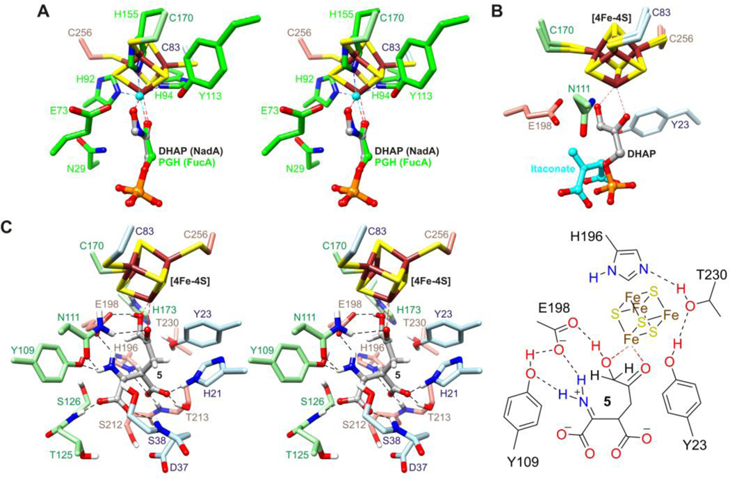
Figure 3.
(A) Stereo diagram of the superimposition of DHAP bound to the [4Fe-4S] cluster in PhNadA onto PGH bound to zinc (cyan) in FucA (green). (B) Superimposition of the [4Fe-4S] cluster with bound DHAP onto the [4Fe-4S] cluster in the itaconate bound structure of PhNadA. (C) Stereo diagram (left) and schematic drawing (right) of intermediate 5 based on the result in panel B, with C1 and Cβ joined, followed by energy minimization with positional restraints applied to the protein atoms.

