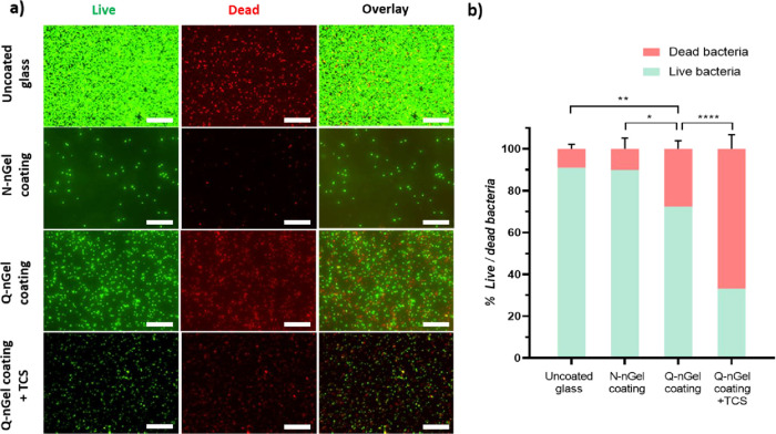
Figure 6.
(a) Fluorescence microscopy images of S. aureus ATCC 12600 that adhered to glass with and without nanogel coatings after 2 h under flow. The scale bar depicts 20 μm. (Intensity has been adjusted for better representation, although for the calculation the originals were used). (b) Adhered bacterial viability was quantified by BacLight LIVE/DEAD staining as a percentage on uncoated glass, N-nGel coating, Q-nGel coating, and Q-nGel coating+TCS. Live and dead bacteria are indicated by green and red, respectively. Experiments were performed on three independent surfaces bearing the nanogel coating and with bacteria that were separately cultured. Differences that are statistically significant are marked with * (p < 0.05), ** (p < 0.01), and **** (p < 0.0001).

