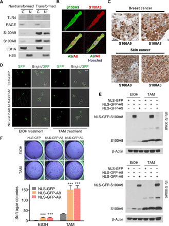
Fig. 2. Nuclear S100A8/A9 and breast transformation.

(A) Western blots of cytosolic (C) and nuclear (N) fractions in nontransformed and transformed cells. LDHA and histone 2B (H2B) are used as subcellular compartment markers. (B) Confocal imaging of S100A8 and S100A9 immunofluorescence costaining in a transformed cell. (C) S100A8 and S100A9 immunohistology from clinical samples of breast cancer (top) and skin cancer (bottom). Representative nuclear localizations of S100A8 or S100A9 are marked by arrows. Scale bar, 50 μm. Data are obtained from The Human Protein Atlas database. (D) Bright-field and GFP fluorescence imaging demonstrate nuclear-specific expression of the indicated derivatives in nontransformed and transformed cells. Scale bar, 50 μm. (E) Western blots of whole-cell lysates show expression of nuclear localization signal (NLS)–GFP–S100A8 (top) and NLS-GFP-S100A9 (bottom). IB, immunoblot. (F) Representative colony formation (top) and quantification (bottom) of soft agar assay with EtOH- or TAM-treated cells expressing indicated protein. Data are means ± SEM; n = 6; ***P < 0.001 compared with corresponding NLS-GFP group; t test. IB, Immunoblot.
