
Fig. 4. Cardiolipin deficit in muscle mitochondria of Hacd1-KO mice is responsible for reduced mitochondrial coupling.
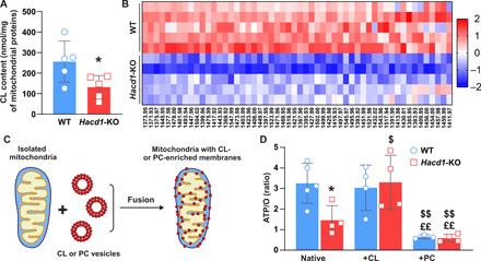
(A) Total cardiolipin content of mitochondria isolated from the tibialis anterior muscle, normalized to the mitochondrial protein content. (B) Heatmap of individually identified cardiolipin species in mitochondria isolated from tibialis anterior muscle of Hacd1-KO mice, compared to WT mice. Colors show row z scores. (C) Diagram depicting the experimental steps allowing to enrich mitochondrial membranes with phospholipids. (D) ATP/O ratio calculated from simultaneous recording of O2 consumption and ATP production of native, cardiolipin-enriched (+CL), and phosphatidylcholine-enriched (+PC) mitochondria isolated from tibialis anterior muscle of WT and Hacd1-KO mice. Error bars ± SEM.; *P < 0.05 versus WT, $P ≤ 0.05 and $$P ≤ 0.01 versus native, and ££P ≤ 0.01 versus +CL.