
Fig. 1. Analysis of Hawaiian stony corals.
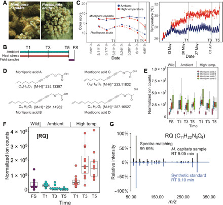
(A) Images of M. capitata and P. acuta from Kāne‘ohe Bay, O‘ahu. Photo credit: D. Bhattacharya, Rutgers University. (B) Experimental design. FS refers to field samples. These are the wild coral individuals that were examined at the end of the thermal stress experiment. (C) Color scores for the (right image) ambient- and high-temperature treated coral species M. capitata and P. acuta at the Hawaiʻi Institute of Marine Biology. Ambient-temperature tanks are shown in variations of red, and high-temperature tanks are shown in variations of blue. The color scores represent a proxy for algal symbiont density in coral holobionts with low values indicating bleaching phenotype. The sharp score decrease for P. acuta under ambient tank conditions is explained by the unexpected warming event that occurred in Kāneʻohe Bay, Oʻahu from which the culture water was drawn. Vertical gray lines indicate sampling points T1 (22 May 2019), T3 (3 June 2019), and T5 (7 June 2019). (D) Structures of MAs identified in the coral holobiont. (E) Accumulation of total MAs in M. capitata over the duration of the tank experiments and from wild populations. (F) Metabolite arginine-glutamine (RQ) in M. capitata changes over time during thermal stress (T1 to T5). (G) The metabolite C11H22N6O4 that showed accumulation under heat stress matches the synthetic standard of RQ dipeptide in retention time and MS2 spectra.