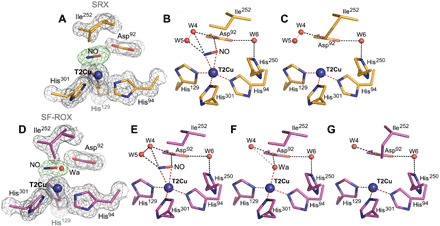
Fig. 4. T2Cu site in NO-bound/T1Cu-reduced Br2DNiR: SRX v SF-ROX structures.

(A) SRX NO-bound structure (1.19 Å; orange) has two conformations of the active site: (B) one with NO present and (C) with three coordinated T2Cu site. (D) In the 1.3 Å XFEL FRIC structure (magenta), there are three alternative conformations of the T2Cu site: (E) one with NO, (F) second with water, and (G) third with three coordinated T2Cu site where NO has vacated the T2Cu site with alternative Ile252 conformation of CD atom flipping down to fill the space. Water molecules are shown as red spheres, and T2Cu is shown as a blue sphere. Distances for possible hydrogen bonding are shown as black dashed lines; gray dashed lines represent unlikely bonding due to steric restraints, and coordination distances to T2Cu are shown as red dashed lines. OMIT Fo − Fc electron density maps around T2Cu ligands are contoured at 5σ level and colored green. Gray 2Fo − Fc electron density map is contoured at 1σ level.
