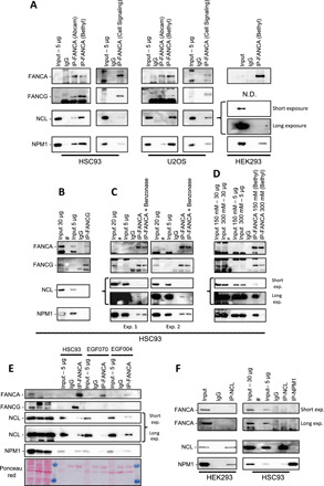
Fig. 4. FANCA immunoprecipitates with NPM1 and NCL.

(A) Anti-FANCA rabbit antibodies from Abcam, Bethyl, and Cell Signaling laboratories were used to immunoprecipitate FANCA in cell extracts from exponentially growing human lymphoblasts (HSC93), human bone osteosarcoma cells (U20S), or human embryonic kidney cells (HEK293). Immunoblotting was performed with anti-FANCA Bethyl or Abcam antibodies and antibodies against FANCG, NCL, and NPM1. N.D., Not Done. (B) Immunoprecipitation (IP) with a FANCG antibody followed by immunoblot with antibodies against the indicated proteins. Different quantities of input fractions were analyzed to clearly visualize FANCA and FANCG. (C and D) Immunocomplexes isolated by the FANCA antibody were treated with benzonase [(C), two independent experiments], 150 nM NaCl, or 300 nM NaCl (D) before immunoblot analysis. Different quantities of input fractions were analyzed to clearly visualize FANCA and FANCG. (E) Immunoprecipitation with a FANCA antibody in cell extracts from HSC93 (WT), EGF070 (FANCG−/−), and EGF004 (FANCG−/−) cells followed by immunoblot with antibodies against the indicated proteins. Different quantities of input fractions were loaded. (F) NCL or NPM1 were immunoprecipitated from HEK293 or HSC93 cells. Immunoblot showing the coimmunoprecipitation of the indicated proteins when cell extracts were immunoprecipitated with anti-NCL or anti-NPM1 antibodies.
