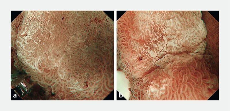
Fig. 2.

The influence of the previous biopsy procedure on magnifying endoscopy with narrow-band imaging (M-NBI) findings in superficial non-ampullary duodenal epithelial tumors. a M-NBI of a duodenal adenoma before biopsy. A clear demarcation line is identified according to distinct differences in the microsurface (MS) pattern between the lesion and the background mucosa. Vessel plus surface (VS) classifications: V, Because of the presence of white opaque substance (WOS), the morphology of the subepithelial microvessels cannot be observed, making this an absent microvascular (MV) pattern. S, The WOS has a regular reticular pattern with a symmetrical distribution and regular arrangement. The marginal crypt epithelium shows a regular arrangement and symmetrical distribution. Thus, this lesion was graded as a regular MS pattern using WOS as a marker for the MS pattern. The VS classification of this lesion was absent MV pattern plus regular MS pattern (WOS +) with a demarcation line. Therefore, the M-NBI diagnosis was non-malignant. b M-NBI of the same lesion after biopsy. The presence of WOS makes it impossible to discern the subepithelial MV pattern of the lesion. Analysis of the WOS morphology shows an irregularly distributed fine WOS, with a variety of morphologies, from speckled to polygonal (irregular WOS). Therefore, the M-NBI diagnosis was cancer. M-NBI findings were compromised by the previous biopsy procedure itself.
