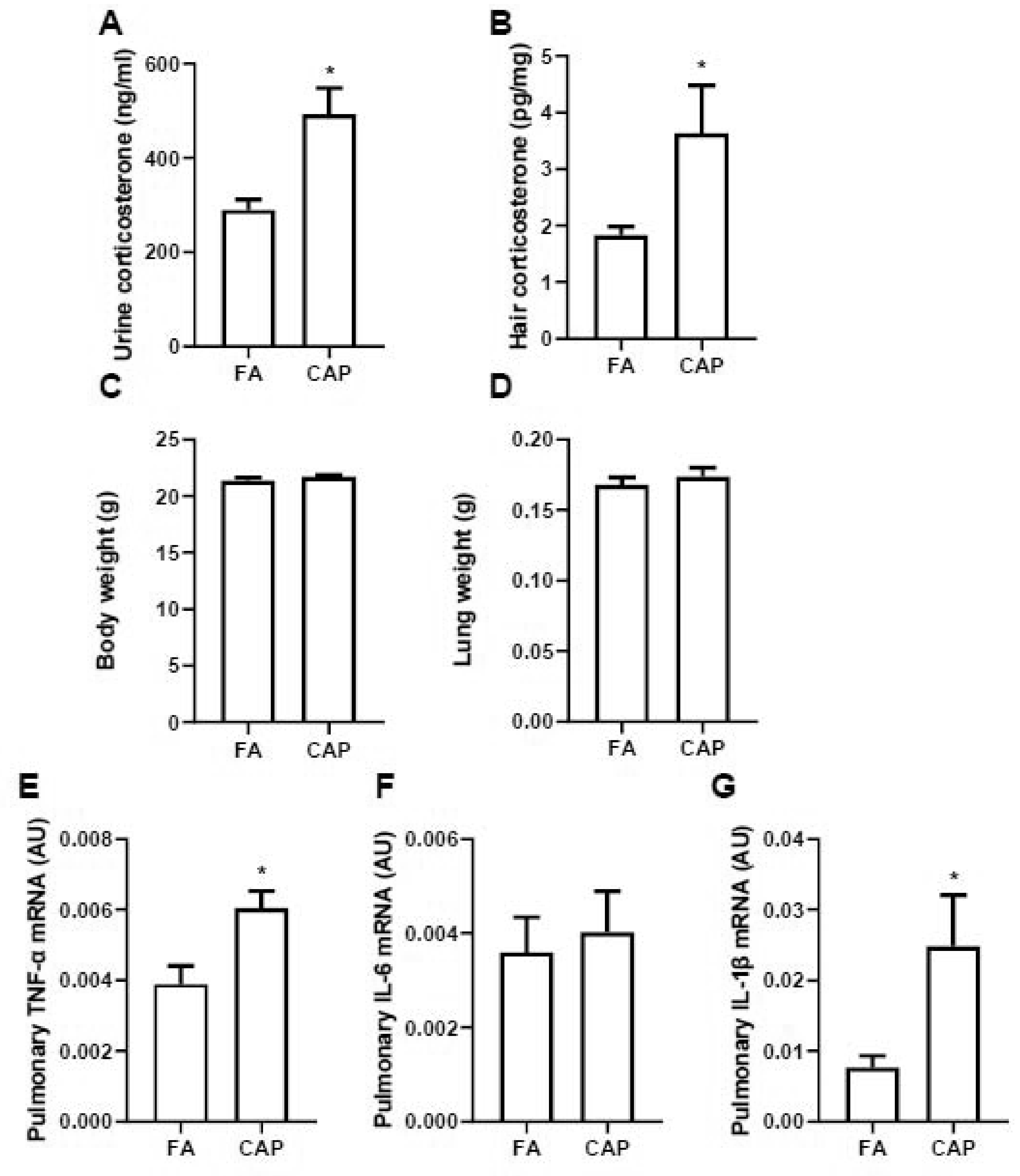
Figure 2. Exposure to ambient PM2.5 activates HPA axis.

A and B, female C57Bl/6J mice were exposed to FA or CAP for 3 months and then the 24-hour urine and hair samples were collected for corticosterone level assessment. Corticosterone levels in urine (A) and hairs (B) were assessed and presented. Their body weight (C) and lung weight (D) were collected and pulmonary inflammatory gene expressions (E, TNF-α; F, IL-6; G, IL-1β) were assessed using real time RT-PCR. n = 10/group. *p<0.05 versus FA, student t test.
