Figure 15.

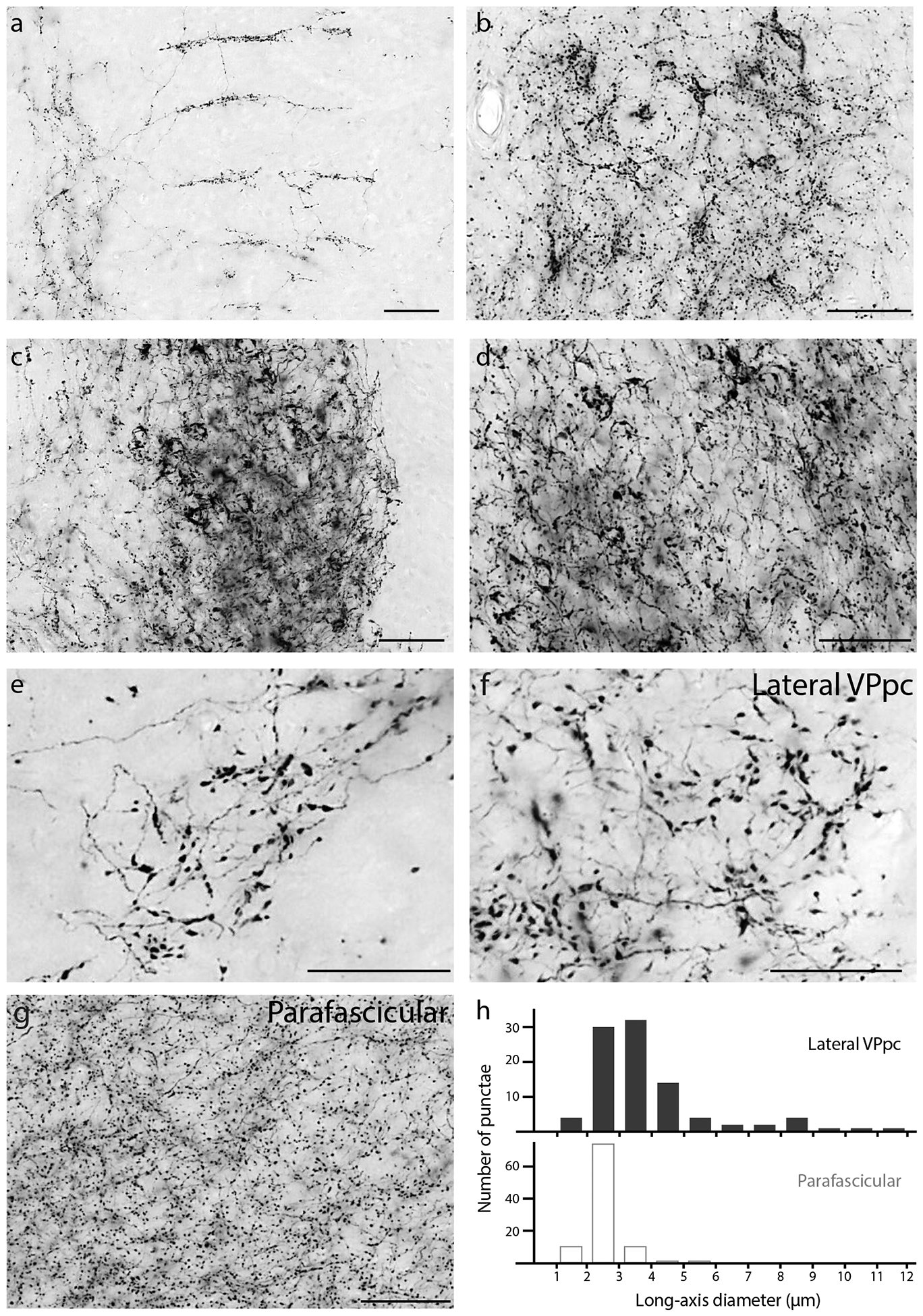
Atypical morphologies of Syp-mCherry-labeled boutons in the: (a) Medial prefrontal cortex (infralimbic area; right side is medial); (b) Septohippocampal nucleus; (c) Oval BST subnucleus; (d) Lateral capsular subdivision of the CeA; (e) PC thalamic nucleus; (f) VPpc (lateral) thalamic nucleus; and (g) PF thalamic nucleus. The large boutons in the PC and lateral VPpc (e–f) form a striking contrast with the uniformly small, grainy appearance of boutons in the neighboring PF (g), which is the typical appearance of PB terminal fields in most other brain regions. (h) The distribution of bouton sizes (long-axis diameter) in the lateral VPpc includes many with larger diameters than in the dorsally adjacent PF.
