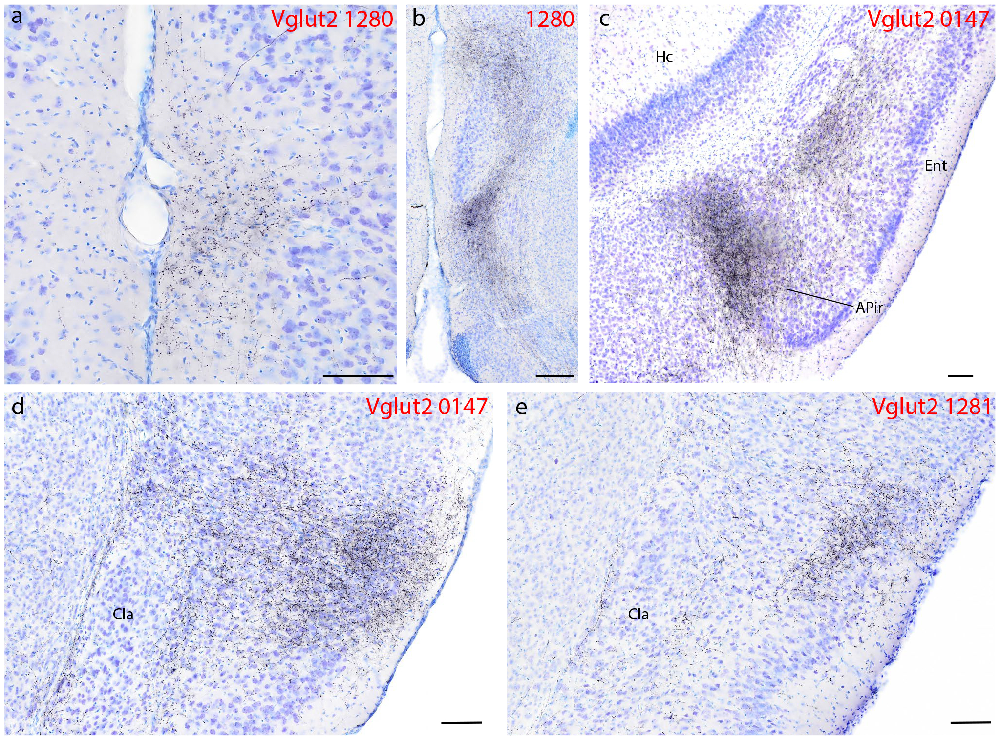
Figure 7.

Syp-mCherry labeling (black, NiDAB) in the cerebral cortex of Vglut2 cases (case numbers at upper-right of each panel). (a) Moderately dense labeling in the medial prefrontal cortex, within the thicker molecular layer (layer 1) of the dorsal peduncular cortex immediately ventral to the infralimbic area (Hurley et al., 1991). (b) Dense labeling in the “septo-olfactory area” aggregates dorsal and ventral to the tenia tecta, which has a thickened layer 2 (Hurley et al., 1991). (c) Dense Syp-mCherry labeling in layer 5 of the rostral entorhinal (Ent) cortex and in the amygdalopiriform transition cortex (APir). (d) Dense glutamatergic Syp-mCherry labeling covers all layers of the mid-insular cortex, but avoids the principal claustrum nucleus (CLA). In one Vglut2 case with an injection site centered in the lateral PB (1281), labeling in the insular cortex clustered superficially, over layer 3. All scale bars are 100 μm. Other abbreviations: Hc, hippocampus.
