
Figure 8.
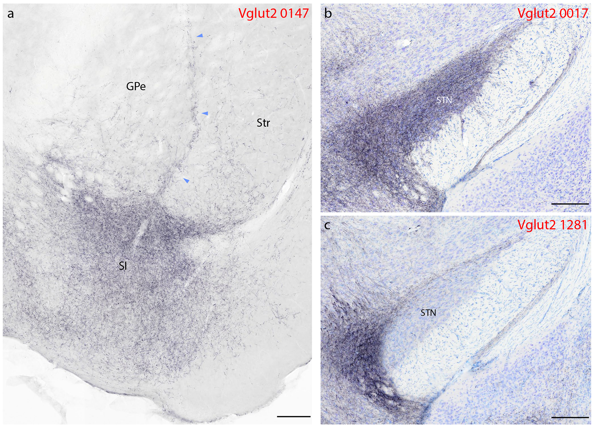
Syp-mCherry labeling in basal forebrain and basal ganglia. (a) In addition to light punctate labeling in both the striatum (Str) and globus pallidus (GPe), a band of Syp-mCherry-labeled boutons extends dorsally between these two structures, up from a dense terminal field in the basal forebrain, primarily within the substantia innominata (SI). (b–c) We found dense labeling in the subthalamic nucleus (STN) in only one case, where the injection site spread rostrally from the PB into the pedunculopontine tegmental area (b, 0017). Vglut2 case 1281 (c) and other Vglut2 cases had little to no labeling in the STN. Other abbreviations: ic, internal capsule; ot, optic tract. Scale bars are 200 μm.