
Figure 9.
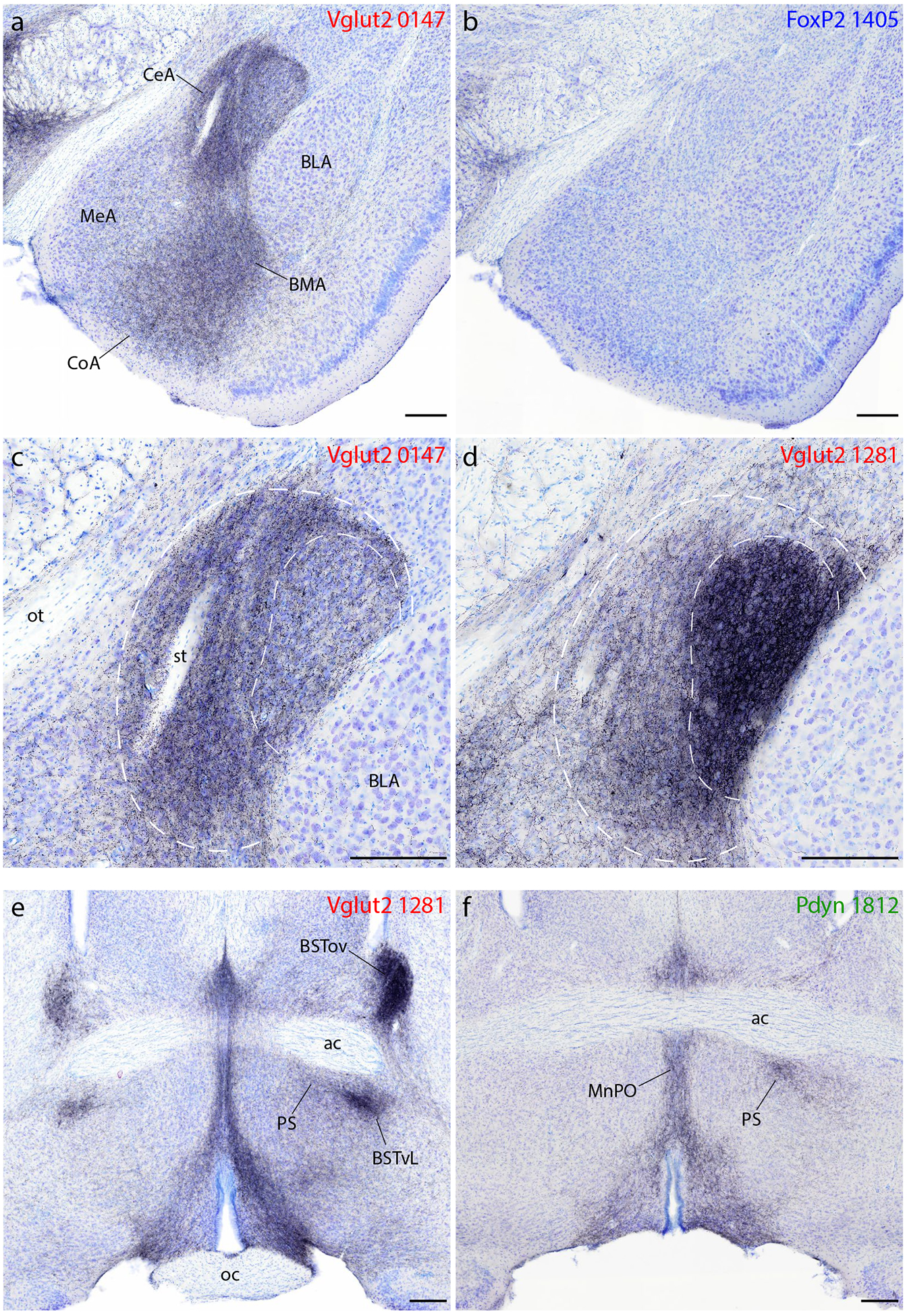
Syp-mCherry labeling in the amygdala, bed nucleus of the stria terminalis (BST), and preoptic area. (a–b) All Vglut2 cases had dense labeling in the central (CeA), basomedial (BMA), and cortical (CoA) nuclei of the amygdala and lighter labeling in the medial nucleus (MeA), with little to no labeling in the basolateral nucleus (BLA). (b) FoxP2 (and Pdyn) cases had virtually no Syp-mCherry labeling in the amygdala. (c–d) Injection site location within the PB affects the distribution of labeling within the CeA. An injection site centered in the medial PB (c, case 0147) produced denser Syp-mCherry labeling in the medial CeA subdivision (outer dashed line), while an injection into the lateral PB (d, case 1281) produced denser labeling in the combined lateral and lateral capsular CeA subdivisions (inner dashed line). (e–f) Vglut2 cases contained dense labeling in the anterior, lateral BST. In this region of the BST, the densest labeling in the oval subnucleus (BSTov) appeared after an injection centered in PBeL (e, case 1281). Vglut2 cases also had consistently heavy labeling in a focal subregion of the ventrolateral BST (BSTvL) and in the adjoining preoptic parastrial nucleus (PS), as well as the median preoptic nucleus (MnPO). (f) Pdyn (and FoxP2) cases had much lighter labeling in the BST, but similarly dense Syp-mCherry labeling in the PS and MnPO. Other abbreviations: ac, anterior commissure; oc, optic chiasm; st, stria terminalis. Scale bars are all 200 μm.