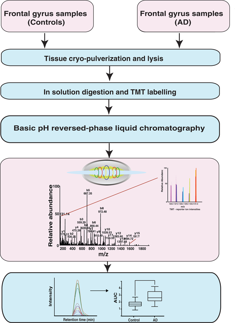
Figure 1.
A schematic of the workflow used to study the proteomic changes in the brain of AD patients. The brain samples were homogenized in liquid nitrogen and lysed in 2% SDS buffer. Brain proteomes from cognitively normal individuals were compared with that of AD patients using a TMT-based quantitative proteomics approach in the discovery step. Validation was carried out using parallel reaction monitoring (PRM) assays for a subset of molecules that were found to be altered in abundance in AD.

