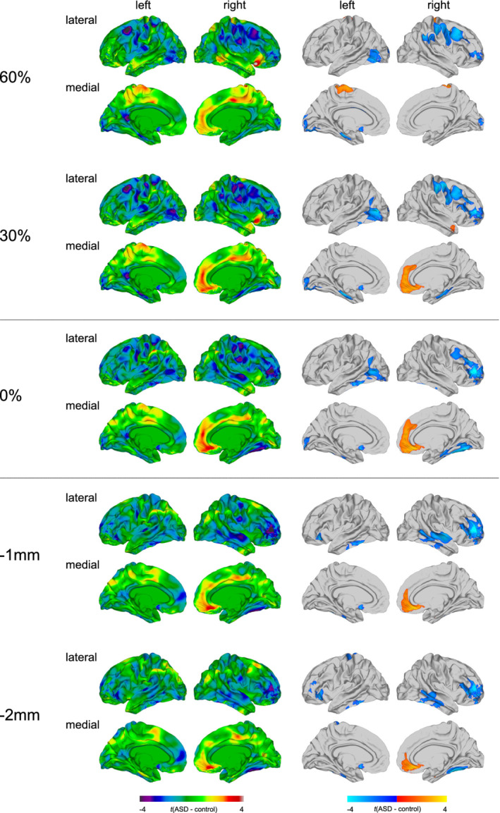
FIGURE 1.

Main effect of group for fractional anisotropy (FA). Regions of increased and decreased FA in individuals with autism spectrum disorder (ASD) compared to typically developing (TD) controls at the gray‐white matter (GWM) boundary (0%), at different cortical thickness (CT) projection fractions within the gray matter (i.e., 30 and 60% CT, sampled from the GWM boundary into the thickness of the cortical ribbon), and within the superficial white matter (sampled at absolute distances of −1 and −2 mm below the GWM boundary). Displayed are the unthresholded (left panel) and thresholded (right panel) t‐maps, where increased FA estimates in ASD are marked in yellow to red (left panel), respectively, red to yellow (right panel), and decreased FA estimates in ASD are marked in cyan to purple (left panel), respectively, blue to cyan (right panel; random field theory [RFT]‐based cluster corrected p < .05, two‐tailed)
