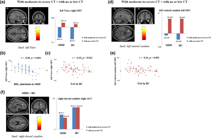
FIGURE 1.

Results of image analyses and correlation analyses. (a) Participants with a history of moderate‐to‐severe CT showed reduced FC between the left NAcc seed and the right OFC compared to participants with no or low level of CT. The altered connectivity between left NAcc and right OFC was significantly correlated with (b) BDI_anhedonia scores in MDD (r = −0.50, p < .001) and (c) physical trait anhedonia scores in HC (r = −0.33, p = .013); (d) Participants with a history of moderate‐to‐severe CT showed decreased FC between the left ventral caudate seed and the left OFC compared to participants with no or low level of CT; (e) The altered connectivity between left ventral caudate and left OFC was significantly correlated with physical trait anhedonia scores in HC (r = −0.41, p = .001); (f) MDD patients had reduced FC between right dorsal caudate and right ACC compared to HC. For image analyses, results were restricted to a mask combining OFC and ACC ROIs. Significance threshold was set at p < .05, FWE cluster level corrected, starting from voxel level p < .001 uncorrected. ACC, anterior cingulate cortex; BDI_anhedonia, sum of anhedonia‐related item 4, 12, and 21 in BDI; CT, childhood trauma; CTQ, childhood trauma questionnaire; FC, functional connectivity; HC, healthy controls; MDD, major depressive disorder; NAcc, nucleus accumbens; OFC, orbital frontal gyrus; PAS, Physical Anhedonia Scale; PSS, Perceived Stress Scale
