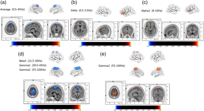
FIGURE 1.

Standardized low‐resolution brain electromagnetic tomography (p < .05). (a) Compared with the control group, the superior frontal cortex (BA 6) of acute tinnitus patients was significantly reduced across the whole frequency band. (b) Compared with the control group, the inferior frontal gyrus (BA 47) of chronic tinnitus patients was significantly enhanced in the delta frequency band. (c) Compared with the control group, the superior temporal gyrus (BA 13) of chronic tinnitus patients was significantly enhanced in the alpha 1 frequency band. (d) Compared with the control group, the superior frontal cortex (BA 6) of chronic tinnitus patients was significantly decreased in the beta 3 and gamma frequency bands. (e) Compared with the chronic tinnitus group, the middle temporal gyrus and parietal gyrus (BA 6, BA 7) of acute tinnitus patients were significantly increased in the gamma frequency band
