Figure 3.
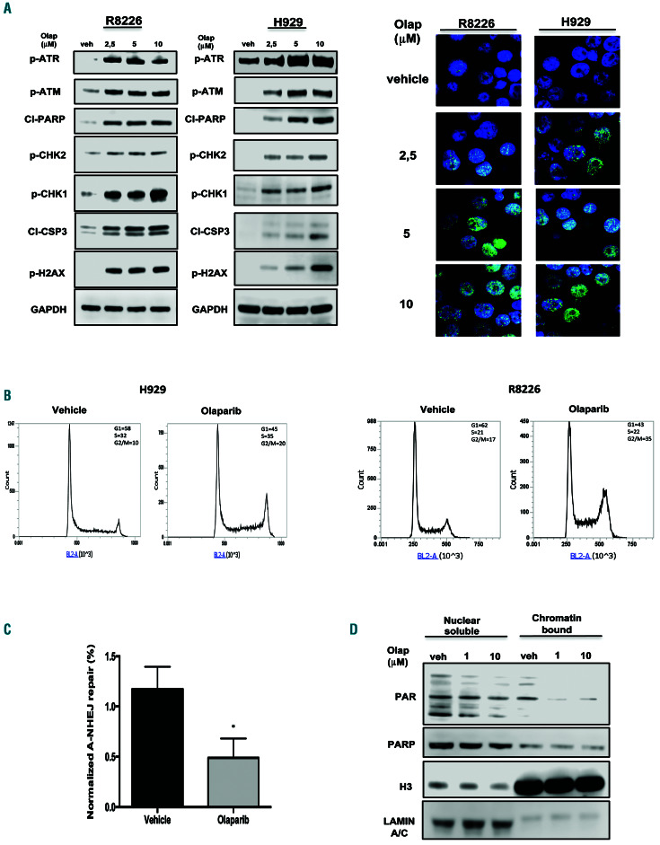
Olaparib activates the DNA damage response in multiple myeloma cells. (A) R8226 and H929 cells were treated with olaparib. Left panel: Immunoblot analysis was performed 24 h after treatment. Right panel: immunofluorescence evaluation by γ-H2AX foci. Representative images of unrepaired double-strand breaks are shown. DAPI (blue) was used for nuclear staining. (B) Cell cycle analysis performed 24 h after treating H929 and R8226 cells with olaparib (2.5 mM). (C) Alternative non-homologous end junction repair was evaluated by EJ2-GFP assay on R8226 cells 48 h af ter treatment with olaparib (2.5 mM) treatment. (D) Western blot analysis of nuclear-soluble and chromatin-bound fractions prepared from R8226 cells. Cells were treated for 30 min with vehicle or olaparib as indicated. Histone H3 and LAMIN A/C were used as positive markers for chromatin and nuclear-soluble fractions, respectively. Data are representative of at least three independent experiments. *P<0.05; **P<0.01. Olap: olaparib; A-NHEJ: alternative non-homologous end junction; PAR: poly (ADP)-ribose; PARP: poly (ADP)-ribose polymerase; H3: histone H3.

