Fig. 4.

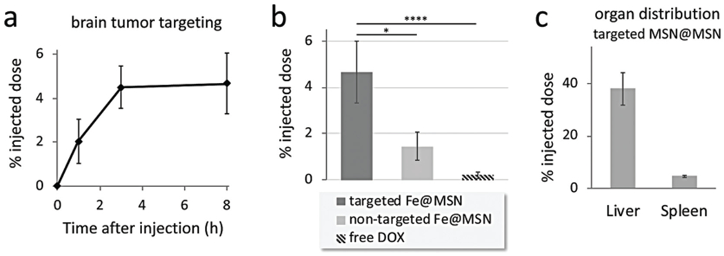
Targeting of CREKA-targeted Fe@MSN nanoparticles to brain tumors. The deposition of CREKA-targeted DOX-loaded Fe@MSN particles in GBM was evaluated in mice bearing orthotopic CNS-1 brain tumors. (a) The accumulation of targeted Fe@MSN particles in brain tumors is shown after 1, 3 and 8 h from i.v. administration at a dose of 5 mg DOX per kg b.w. (n = 5 mice in each group). (b) The accumulation of targeted Fe@MSN in brain tumors was compared to non-targeted Fe@MSN or free DOX using a dose of 5 mg kg−1 DOX for all formulations. Grouped analysis ANOVA; correct for multiple comparisons using the Holm–Sidak method. P values: * <0.01, **** <0.0001. (c) The accumulation of targeted Fe@MSN in liver and spleen is shown after 8 h from i.v. administration at a dose of 5 mg kg−1 DOX (n = 5 mice in each group).
