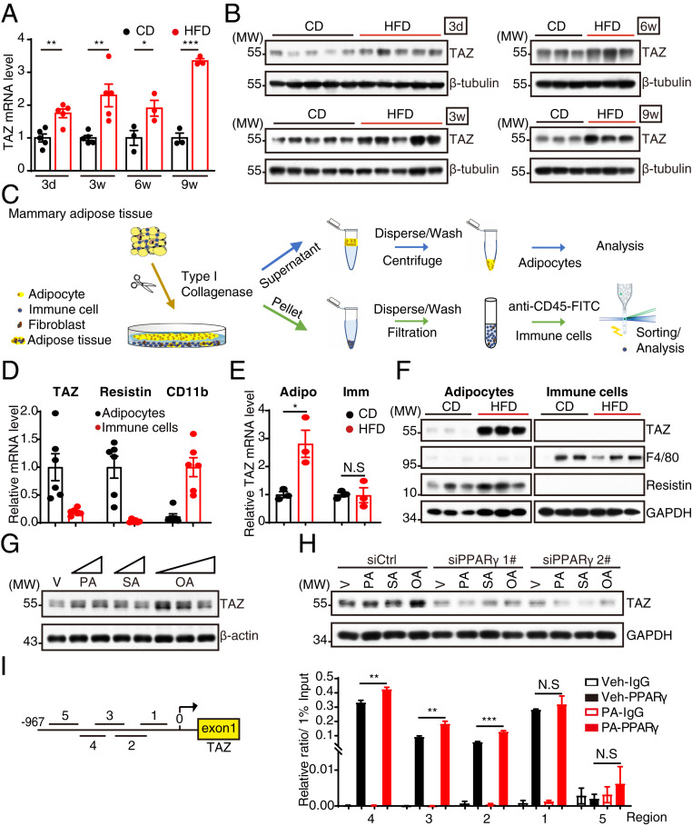
Fig. 1.
Diet-induced adiposity induces TAZ expression in adipocytes through the FFA/PPARγ axis. (A and B) Analysis of TAZ expression in MAT fed with CD or HFD for indicated times by RT-qPCR (A) and Western blot (B). (C) Trial schematic for isolation of mature adipocytes and immune cells from MAT. (D) RT-qPCR analysis of TAZ, Resistin, and CD11b expression in adipocytes and immune cells isolated from MAT. (E and F) RT-qPCR (E) and Western blot (F) analysis of gene or protein expression in adipocytes and immune cells isolated from MAT after feeding CD or HFD for 1 wk. (G) The 3T3-L1 adipocytes were treated with FFAs (PA, 100 and 200 μM; SA, 100 and 200 μM; OA, 100, 200, and 400 μM) or vehicle for 12 h and then harvested for Western blot analysis. (H) The 3T3-L1 adipocytes were transfected with small interfering RNA (siRNA) targeting PPARγ; 48 h later, the cells were treated with 400 μM FFAs for another 12 h. The cells were then harvested for Western blot analysis. (I) The 3T3-L1 adipocytes were treated with 400 μM palmitic acid or vehicle for 12 h and then lysed for ChIP analysis with antibody against PPARγ. Five regions from −967 ∼ −39 bp (base pair) were examined by RT-qPCR for PPARγ binding affinity. MW, molecular weight; Adipo, adipocyte; Imm, immune cell; V and Veh, vehicle. Data shown are mean ± SEM. Data were analyzed using Student’s t test (A and E) and two-way ANOVA (I). N.S.: no significance, *P < 0.05, **P < 0.01, ***P < 0.001.

