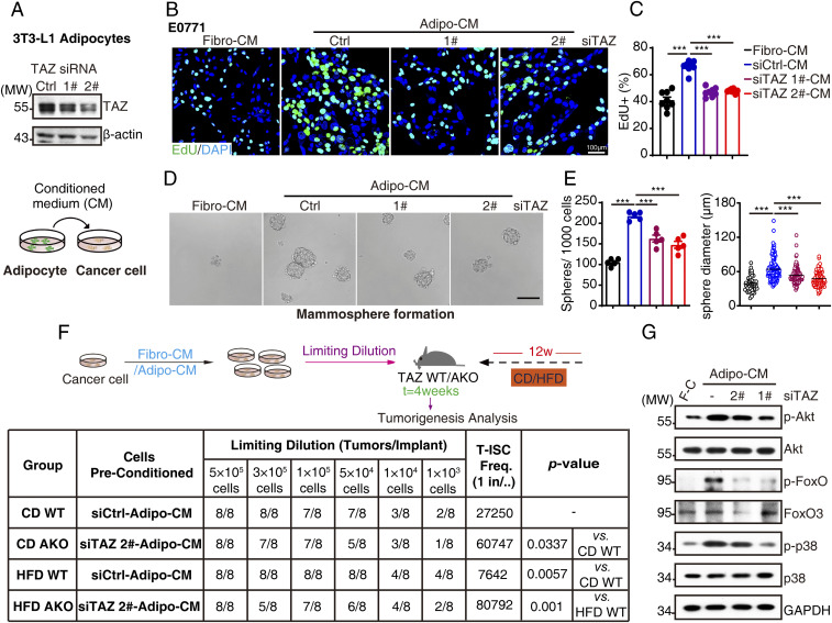
Fig. 2.
Adipocytic TAZ promotes breast cancer proliferation and stemness. (A) The 3T3-L1 adipocytes were transfected with siRNA targeting TAZ; 72 h after transfection, TAZ knockdown efficiency was analyzed by Western blot (Top); schematic representation of Adipo-CM and breast cancer cell coculture assay (Bottom). (B and C) E0771 cells were cultured in control or TAZ knockdown–Adipo-CM for 72 h and subjected to EdU incorporation assays. The new generation cells were detected via EdU (green). 4′,6-diamidino-2-phenylindole (DAPI)-stained nuclei are in blue. Merged view of EdU (green) and DAPI (blue) showing the overlap (B); the quantification for EdU staining was shown (C). (D and E) One thousand E0771 cells were suspension-cultured in mammosphere formation medium containing control or TAZ knockdown–Adipo-CM or Fibro-CM for 10 d; the representative images were obtained by microscopy (scale bar, 100 μm) (D). Mammosphere numbers were counted, and the diameter of mammosphere was measured by Image J software and shown (E). (F) E0771 cells were washed, incubated with control or TAZ knockdown–Adipo-CM for 30 min, and then harvested for Western blot analysis with indicated antibodies. (G) Trial schematic for breast cancer cell culture with different CMs and strategies for limit dilution analysis (Top). Eight weeks after culture with specified CM, E0771 cells were injected into TAZ WT or AKO mice mammary fat pad with indicated numbers (limit dilution); 4 wk after injection, stem cell frequency was determined (n = 8). F-C for Fibro-CM, A-C for Adipo-CM, T-ISC for tumor-initiating stem cell (Bottom). Data shown are mean ± SEM. Data were analyzed using one-way ANOVA. ***P < 0.001.

