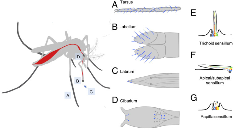
Fig. 4.
Anatomy of mosquito taste organs and sensilla. (Left) Illustration of mosquito with taste organs highlighted in dark gray. (A–D) Taste sensilla are depicted in blue: (A) tarsus (third to fifth segments), (B) labellum, (C) apical/subapical portion of labrum, and (D) cibarium. (E–G) Morphology of taste sensilla containing several gustatory receptor neurons (in color) and one mechanosensory neuron (in dark gray): (E) trichoid sensillum, (F) apical/subapical sensillum, and (G) papilla sensillum.

