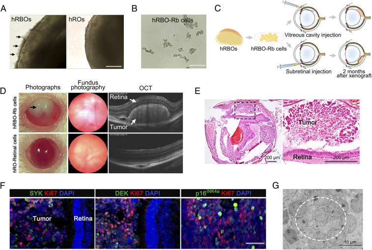
Fig. 3.
Orthotopic engraftment of hRBOs in NSG mice. (A) Representative bright-field image of 120-d-old hRBOs and hROs. Arrows indicate excessive outgrowth of Rb cells, subsequently leading to detachment from the organoids. (Scale bar: 400 µm.) (B) Rb cells derived from hRBOs (hRBO-Rb cells) can be maintained in long-term culture in suspension. (Scale bar: 200 µm.) (C) Schematic view of the procedure for generating tumor engraftments in NSG mice. (D) Ophthalmic examination of mouse eyes after subretinal engraftment. Representative photograph of engrafted eyes at 2 mo after subretinal injection (Left). hRBO-Rb cell-engrafted eye showing leukocoria-like features (black arrow). Representative fundus photography and OCT images of an hRBO-Rb cell-engrafted eye showing an obvious Rb tumor in the retina. Retinal cells derived from hROs (hRO-retinal cells) served as normal controls (Bottom). (E) Representative H&E staining of hRBO-Rb cell-engrafted eyes at 2 mo after subretinal injection. The boxed region is magnified on the right. (Scale bar: 200 µm.) (F) Immunostaining of Rb signature genes Ki67, SYK, DEK, and p16INK4a in hRBO-Rb cell-engrafted eyes at 3 mo after subretinal injection. (Scale bar: 50 µm.) (G) Representative TEM image of Rb engraftment. The dashed line denotes the typical rosette structure. (Scale bar: 10 µm.)

