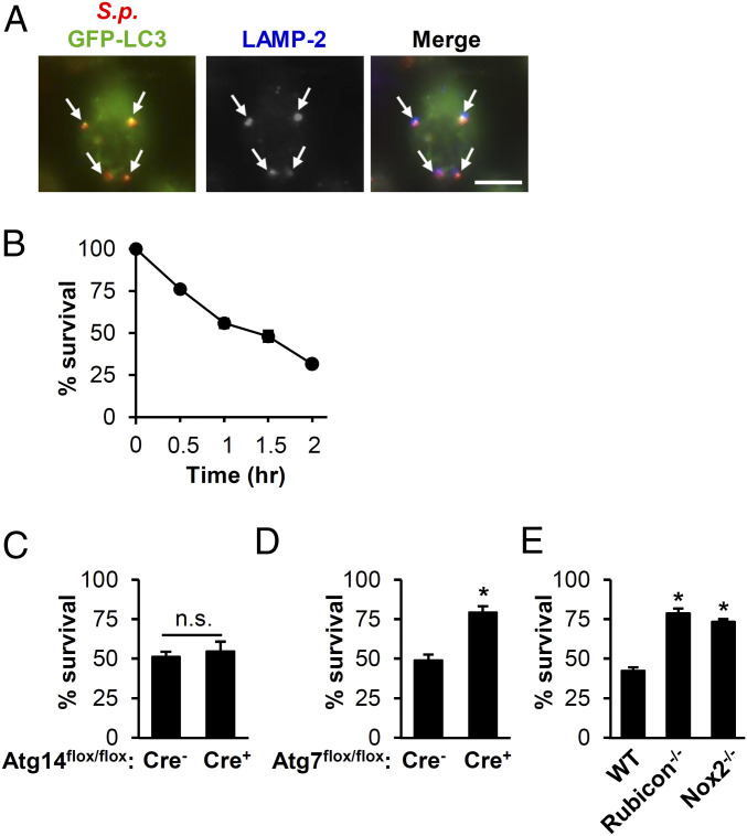
Fig. 4.
LAP contributes to clearance of S.p. by BMDMs. (A) BMDMs transfected with GFP-LC3 were infected with S.p. At 1-h postinfection, cells were stained for S.p., LC3, and LAMP-2. Shown are images representative of three independent experiments. Arrows: S.p. colocalized with LC3 and LAMP-2. (Scale bar, 5 µm.) (B) Survival of S.p. in BMDMs from young WT mice. Data show the mean ± SEM of a representative experiment from, at least, three independent experiments. (C–E) The percent S.p. survival was determined after 1.5-h infection of BMDMs from Atg14-KO (Atg14flox/flox-LysM-Cre+) or control (Atg14flox/flox-LysM-Cre−) mice (C); Atg7-KO (Atg7flox/flox-LysM-Cre+) or control (Atg7lox/flox-LysM-Cre−) (D); WT, Rubicon−/−, or Nox2−/− (E). Data show mean ± SEM from a representative experiment from, at least, two independent experiments. *, P < 0.05 by the Mann–Whitney U test. n.s., not significant; S.p., Streptococcus pneumoniae.

