Fig. 3.
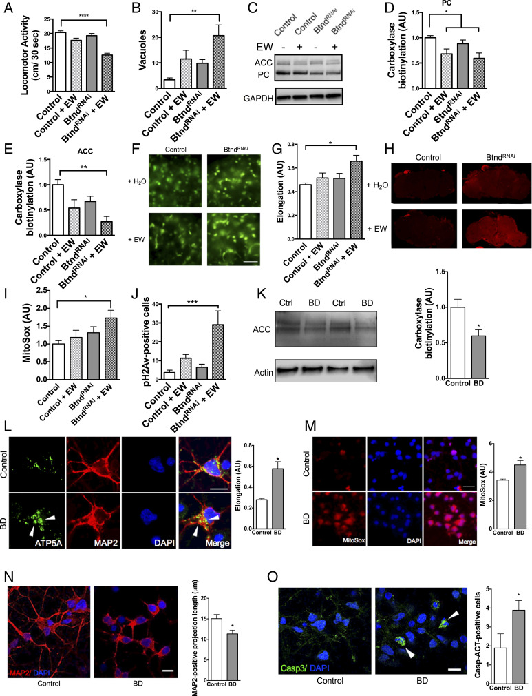
Biotin deficiency alone is capable of causing neurodegeneration and mitochondrial dysfunction in Drosophila and differentiated human cortical neurons. Biotin-deficient flies were generated by treating BtndRNAi flies with an egg white (avidin)-supplemented diet (+EW). Biotin-deficient flies show behavioral deficits (A), increased markers of neurodegeneration (B), and a loss of carboxylase biotinylation compared to control flies (C–E). (F–I) Biotin-deficient flies show abnormal mitochondrial morphology and function. GFP-tagged mitochondria were analyzed using the Mito ImageJ Plugin (F and G). (Scale bars, 5 μm.) Oxidative stress was indicated using MitoSox dye and quantification from confocal Z-stacks (H and I). (J) Biotin deficiency causes DNA damage as shown by pH2Av-positive foci in the brains of avidin-fed flies. Fly feeding experiments were analyzed by two-way ANOVA with Tukey’s post test; n = 6 per genotype. Control genotype (A–E, H–J) was elav-GAL4/+. Control genotype (F and G) was elav-GAL4/+;UAS-mito-GFP/+. (K–M) Similarly, cortically differentiated iPSCs treated with biotin-deficient (BD) media showed a reduction in carboxylase biotinylation (K), abnormal mitochondrial morphology as shown by ATP5A immunolabeling (L, arrowheads referencing enlarged mitochondria), and an accumulation of superoxide oxidative stress by MitoSox dye (M). Cells treated with biotin-deficient media also showed shortened MAP2-positive projection length (N) and increased caspase activation (O, arrowheads refe1rencing activated caspase-positive cells). (Scale bars = 5 μm.) Cell culture experiments were analyzed by t test. *P < 0.05, **P < 0.01, ***P < 0.001, ****P < 0.0001.

