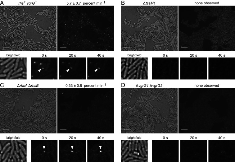
Fig. 3.
Rhs promotes T6SS-1 sheath assembly. Time-lapse fluorescence microscopy of (A) rhs+ vgrG+, (B) ∆tssM, (C) ∆rhsA ∆rhsB, and (D) ∆vgrG1 ∆vgrG strains that express the tssB-gfp fusion. Representative bright-field (Left) and fluorescence (Right) images are presented for each background. (Scale bars, 5 µm.) Time-lapse micrographs are shown below, with white carets indicating coupled assembly−contraction events. T6SS-1 firing frequency is expressed as the number of assembly−contraction events per 100 cells per min. Data are reported as the average ± SEM for at least three independent experiments.

