Fig. 4.
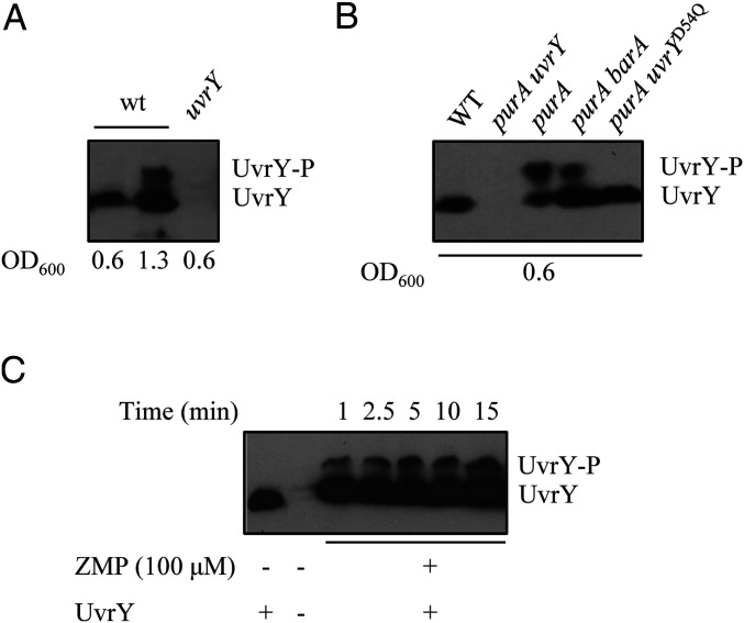
UvrY and UvrY-P detection by Phos-Tag SDS/PAGE followed by Western blot analysis. (A) Lanes 1 and 2, KSB837 (WT); lane 3, IFC6003 (uvrY−). In lanes 1 and 3, cell extracts were obtained from cultures at an OD600nm ≈ 0.6 (nonstimulatory conditions), and, in lane 2, cell extracts were obtained from a culture grown to an OD600nm ≈ 1.3 (stimulatory conditions). (B) Lane 1, KSB837 (WT); lane 2, IFC6005 (uvrY− purA−); lane 3, IFC6003 (purA−); lane 4, IFC6004 (barA− purA−); and lane 5, IFC6005 (uvrY− purA−) harboring plasmid pMX577 (expressing UvrYD54Q). Cell extracts were obtained from cultures at an OD600nm ≈ 0.6 (nonstimulatory conditions). (C) Purified His6-UvrY was incubated in a 40-µL reaction mixture with 100 µM ZMP, and, at the indicated time intervals, 5-µL samples were withdrawn and subjected to Phos-Tag SDS/PAGE followed by Western blot analysis.

