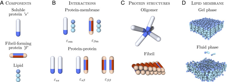
Fig. 1.
Simulation model for membrane-driven fibril formation. (A) Proteins can exist in two distinct conformations: a soluble “” and a -sheet-prone conformation. Lipid molecules are modeled by one hydrophilic head and two hydrophobic tail beads. (B) Protein–membrane and protein–protein interactions present in the system and the associated interaction parameters. (C) Soluble proteins can form oligomers via their tip-to-tip interactions. Protein molecules in the -sheet-forming conformation can assemble into fibrillar structures through the interactions of the blue side patches. (D) The lipid membrane can exist in different structural phases depending on the interlipid interactions set in the model. Shown here are the gel phase ( = 0.775) and fluid phase ( = 1.135).

