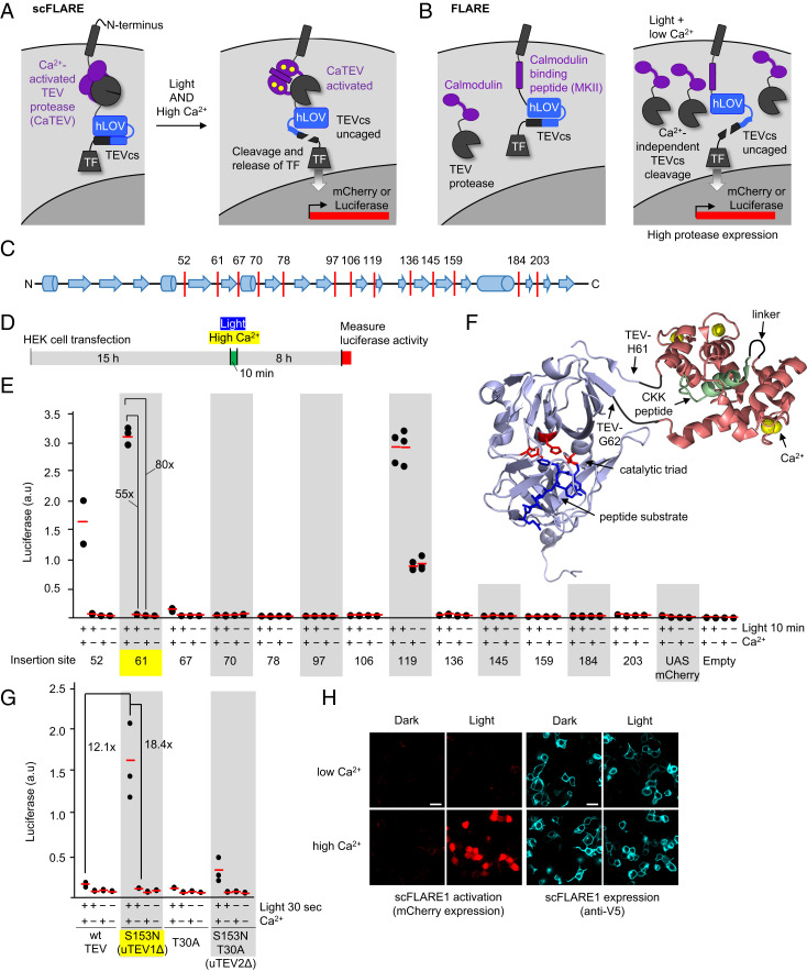
Fig. 1.
Engineering of CaTEV and incorporation into scFLARE. (A) Schematic of scFLARE. From N to C terminus, scFLARE consists of a transmembrane helix, a CaTEV, an LOV domain, a TEV cleavable site (TEVcs), and a TF. High Ca2+ activates the protease, while light alters the conformation of LOV to expose the TEVcs for cleavage. Both light and calcium must arrive coincidentally to enable proteolytic release of the TF, which translocates to the nucleus to drive expression of the reporter gene of choice. (B) The previous two-component FLARE (6, 16) gives background at high protease expression levels. When the FLARE protease copy number is high, TEVcs cleavage can occur under the light + low-Ca2+ condition because of proximity-independent recognition of TEVcs by TEV protease. (C) The domain structure of TEV protease (from Protein Data Bank (PDB) ID: 1LVM) showing loop regions we targeted for insertion (red lines) of Ca2+-sensing modules. (D) The protocol for screening CaTEV variants in the context of scFLARE in HEK 293T cells. Ca2+ was elevated by the addition of 6 mM CaCl2 in the presence of 2 μM ionomycin. Blue light (467 nm) was delivered at 60 mW/cm2 and 33% duty cycle (2 s of light every 6 s). (E) The screening results from D. Calmodulin-CKK was inserted after the indicated residue in TEV, within the context of scFLARE as shown in A. This experiment was performed once with three technical replicates per condition (red lines, mean). (F) Model of the best CaTEV design based on crystal structures of wild-type TEV protease (PDB ID: 1LVM) and calmodulin:CKK complex (PDB ID: 1CKK). The CaM-CKK fusion is inserted between His-61 and Gly-62 of TEV protease. (G) The incorporation of ultraTEV mutations (16) into scFLARE. The same assay as in E, but the light + Ca2+ stimulation time was only 30 s. This experiment was performed once with three technical replicates per condition. (H) Confocal imaging of scFLARE activity in HEK293T cells. scFLARE1 (insertion site 61) was expressed with UAS:mCherry reporter and stimulated for 30 s with blue light and CaCl2 + ionomycin. Cells were fixed, permeabilized, and stained with anti-V5 antibody 8 h later to detect scFLARE expression (scale bars, 20 μm.) This experiment was repeated two times.

