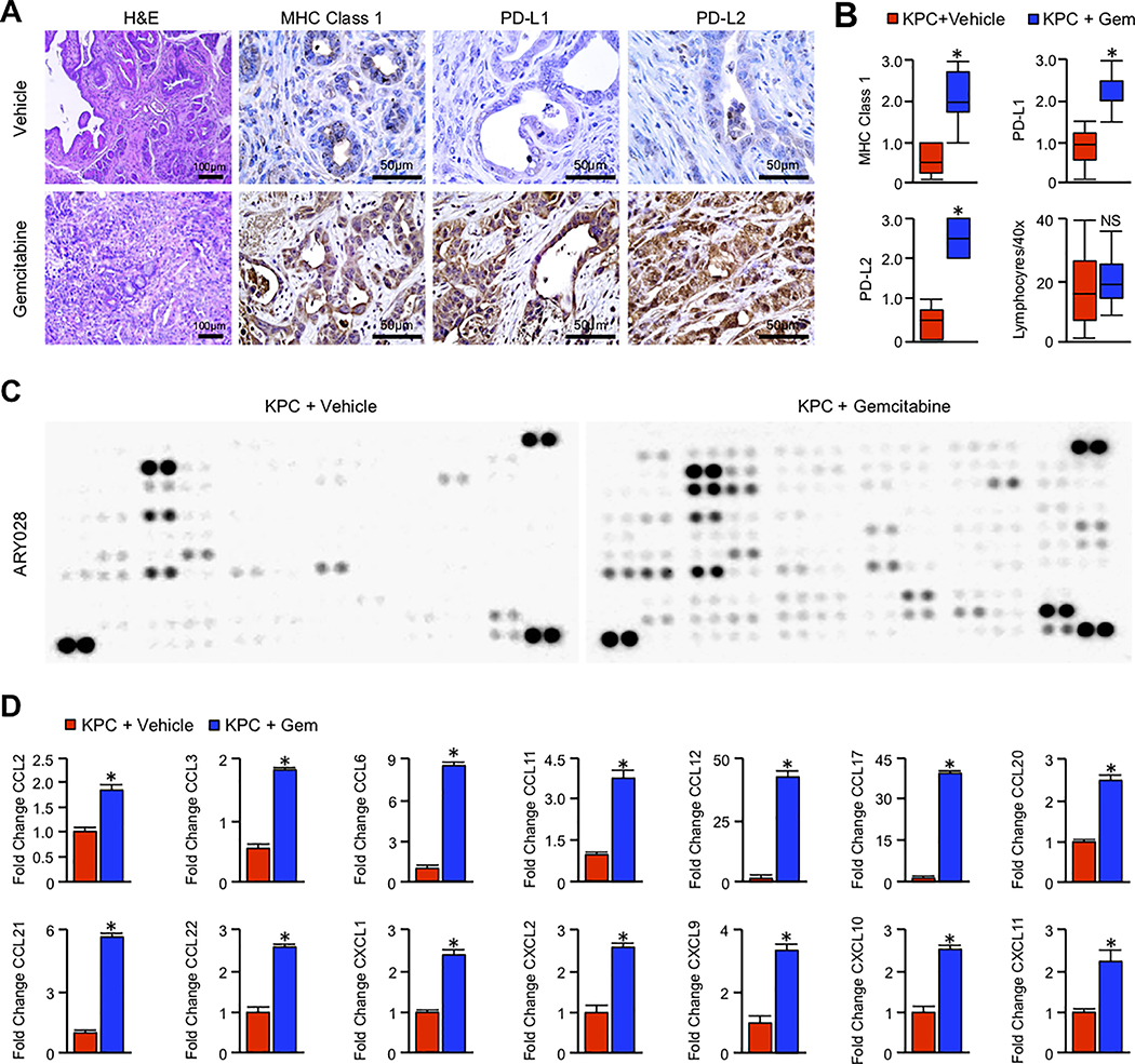
Figure 1. Long-term Gemcitabine treatment alters the immune landscape of murine PDAC.
(A) Pdx1-Cre x LSL-KrasG12D x LSL-TP53R172H (KPC) mice were generated as a model of advanced PDAC. Starting at 90 days (~13 weeks) of age, mice were administered twice-weekly intraperitoneal injections of either PBS vehicle or 100mg/kg Gemcitabine. Pancreas tissues were collected when the animals were moribund. Tissues from vehicle and Gemcitabine treated mice were then stained with H&E or by immunohistochemistry for MHC Class 1, PD-L1, or PD-L2. (B) Tissue sections were evaluated by three blinded investigators. For IHC images, slides were assigned a score by each investigator from 0–3+ based on staining intensity, and composite values displayed as a box plot. For tumor infiltrating lymphocytes, each investigator quantified four H&E stained 40X fields per animal. These values were averaged and represented by box plot (*p < 0.05, N=4/group). (C) Tissues were homogenized and 200μg of tumor lysate evaluated by a high throughput proteome profiler array (ARY028). Representative blots from each group are displayed above. (D) Pixel density was evaluated using ImageJ, and samples normalized to the mean intensity of the reference spots for each blot minus the background density. Composite normalized values for Gemcitabine treated mice were divided by those for vehicle treated mice, and are presented as fold change (*p < 0.05).

